
Figures & data
Figure 1. Preoperative x-ray showing the tumor encompassing the entire third right metacarpal including the metacarpal head (a). Follow-up x-rays one year after metacarpal reconstruction with MFC bone flap in dorsopalmar (b) and lateral view (c). Note the preservation of metacarpal length and distal bone remodeling at the metacarpal head.
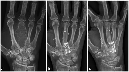
Figure 2. A sterile 3D printed model with a handlebar virtually designed to perfectly fit in the metacarpal defect after tumor resection was used for bone flap elevation. Corticocancellous MFC bone flap after elevation (b).
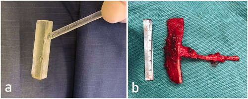
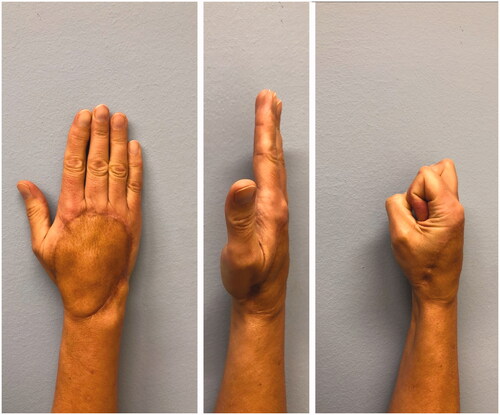
Figure 3. Photograph of the affected hand in two planes two years postoperatively.
Supplemental material