Figures & data
Figure 1. (A) Anastomosis of the latissimus dorsi (LD) flap to the blood vessel on the healthy side. (B) External fixation of both lower limbs.
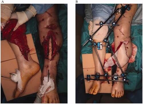
Figure 2. (A) Delay using a tourniquet on the femur of the healthy side. (B) Indocyanine-green angiography of the flap with blood circulation blocked from the pedicle on the healthy side. (C) The oxygen saturation of the flap is measured by near-infrared spectroscopy during delay with a tourniquet.
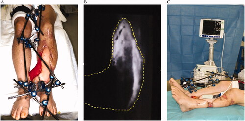
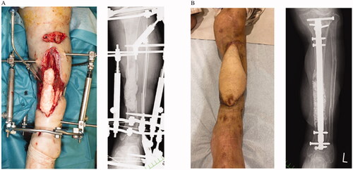
Figure 3. Case 1: (A) Preoperative clinical and radiographic findings. (B) Postoperative findings at 1 year.
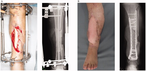
Figure 4. Case 2: (A) Preoperative clinical and radiographic findings. (B) Postoperative findings at six months.