Figures & data
Figure 1. (A) The defect after debridement. (B) Flap design. The blue dot marks the perforator identified with the handled doppler probe.
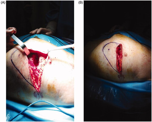
Figure 2. Flap raising. After perforator identification, incision of the lower margin and flap raising (A). (B) De-epithelialization and transposition of the proximal portion of the flap. (C) The wound 1 week post-op.
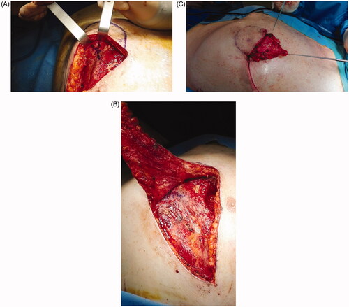
Figure 3. At one month post-op the flap was completely consolidated with the thoracic tissues, scars were well healed and no distortion of the thoracic surface was noted.
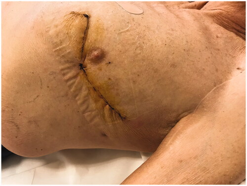
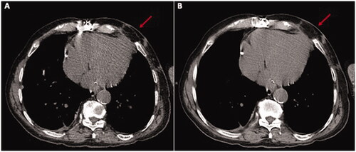
Figure 4. A CT scan executed 1 month post-op (A) and 1 year post-op (B) clearly shows the trophism of the flap (red arrow) and no collection.