Figures & data
Figure 1. Presentation at diagnosis. (A) Right lower extremity. (B) Left lower extremity (medial thigh). (C) Left lower extremity (outer thigh). (D) Left upper extremity. (E) Right upper extremity.
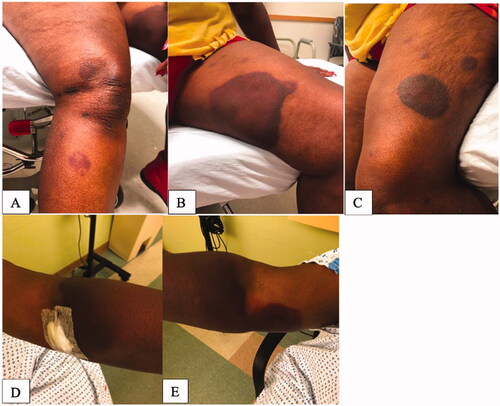
Figure 2. Presentation day of fasciotomy. (A) Right upper extremity with fingers held in flexion.
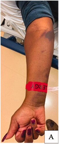
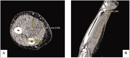
Figure 3. CT Right upper extremity with IV contrast. (A) Axial CT demonstrating a hypodense nonenhancing intramuscular collection within the volar compartment measuring at least 12.3 × 3 × 3 cm. (B) Coronal view of intramuscular collection.