Figures & data
Table 1. Patients’ data.
Figure 1. Images prior to reoperation. (A) MRI T1 weighted image. (B) MRI T1 weighted image with gadolinium enhancement. The dotted line indicates the extent of tumor resection. MRI: Magnetic resonance imaging.
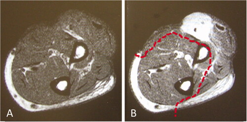
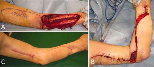
Figure 2. Intraoperative photographs of the patient in Case 1. (A) After extensive resection of the tumor, a large soft tissue defect, which exposed the radius, was observed. (B) While preserving the radial nerve, a 19 × 6.5 cm reverse lateral upper arm flap was raised. (C) The radius was covered with a flap, and a mesh skin graft was performed on a portion of the skin where the forearm muscle was exposed.
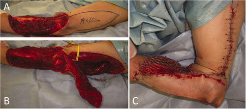
Figure 3. Case 1: Range of motion in the forearm and elbow 8 years and 3 months after the operation. (A) Degree of extension. (B) Degree of flexion.
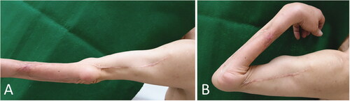
Figure 4. Radiographs of the patient in Case 2. (A) Radiograph taken immediately after the injury. The arm and elbow were contaminated with several windshield pieces. (B) Radiograph taken after debridement. The white arrow indicates deficient humeral condyles, while the black arrow indicates radial head defects.
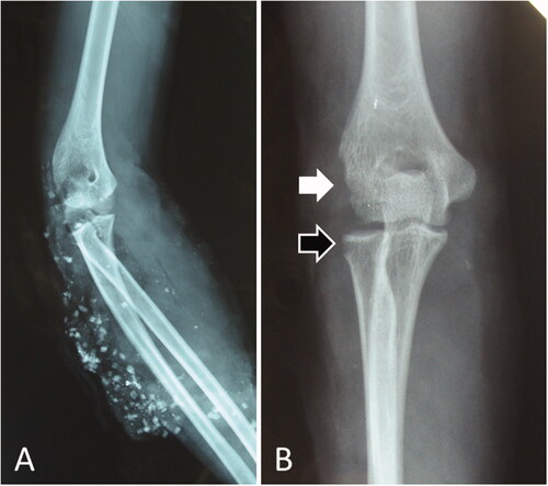
Figure 5. Intraoperative photographs of the patient in Case 2. (A) A 20 × 7 cm reverse lateral upper arm flap was designed. (B) The donor site was closed primarily. (C) One year and 5 months after flap implantation.
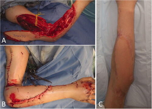
Figure 6. Intraoperative photographs of the patient in Case 3. (A) A 17 × 7 cm reverse lateral upper arm flap was designed. (B) The forearm and the elbow at the time of primary closure of the donor site. (C) Five months after flap implantation.