Figures & data
Figure 1. Left elbow defect following wide local excision of a Merkel cell carcinoma lesion.
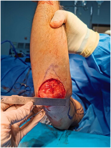
Figure 2. Reverse lateral arm flap design along the axis of the lateral intermuscular septum, extending from the deltoid insertion to the lateral epicondyle. (*) Audible doppler signal of the targeted perforating vessels.
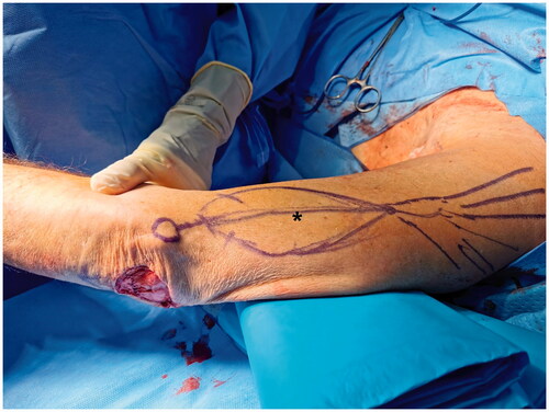
Figure 3. Reverse lateral arm flap pedicle dissection (Arrow) and sparing of the radial nerve (*) and posterior cutaneous nerve of the arm (**).
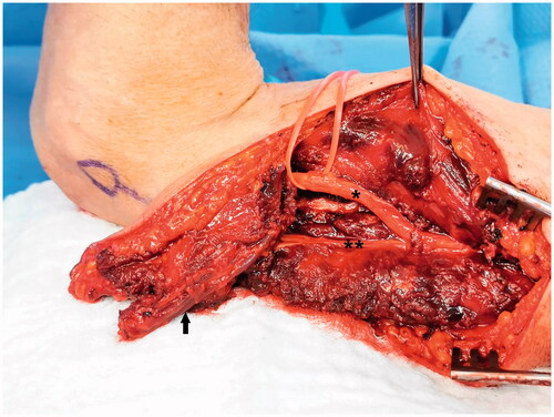
Figure 4. Reverse lateral arm flap rotation and inset.
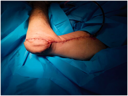
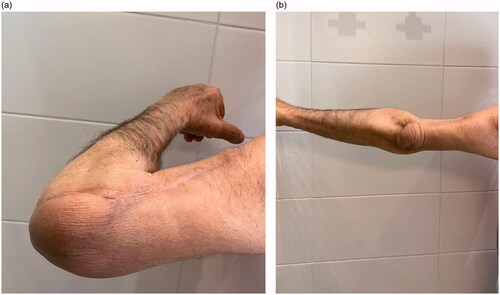
Figure 5. Recovery of elbow range of motion was shown at six months follow-up. (a) Full flexion (b) Full extension.