Figures & data
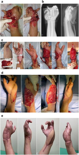
Figure 1. Patient 1. (A) Upon arrival at the emergency room, the blood circulation in the right foot was poor. (B) Multiple fractures and dislocations were observed after performing radiographic examination. (C) The recipient artery was interposed with T portion and the flow was sutured through anastomosis to the TP artery. (D) On day 9, free LD flap was performed. Abbreviation: (E) After the third tangential excision (4 weeks after LD flap). (F) Appearance at 3.5 months after injury. TP: tibialis posterior artery; SSA: subscapular artery; CSA: circumflex scapular artery; TDA: thoracodorsal artery; LD: latissimus dorsi.
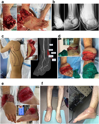
Figure 2. (A) Appearance on his arrival. (B) Radiographic findings show ankle dislocation fracture. (C) Free LD flap was performed at 4 weeks after injury. The recipient artery was interposed with T portion and the flow was sutured through anastomosis to the TP artery. (D) The fourth tangential excision was performed at 3.5 weeks after free LD flap. (E) Findings at 3 months after free LD flap. TP: tibialis posterior artery; SSA: subscapular artery; CSA: circumflex scapular artery; TDA: thoracodorsal artery; LD: latissimus dorsi; LD: latissimus dorsi TDA: thoracodorsal artery; UA: ulnar artery.
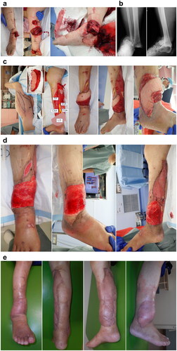
Figure 3. (A) Appearance on his arrival. (B) Radiographic examination on his arrival. (C) Free LD flap was performed at 1 week after injury. The recipient artery was sutured end-to-end to the ulnar artery. (D) After the third tangential excision, (E) Findings at 3 months after free LD flap. LD: latissimus dorsi.