Figures & data
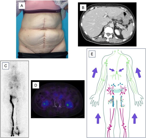
Figure 1. Preoperative findings. Abdominal distension is observed (A). Contrast-enhanced CT shows a large amount of ascites (B). The schema indicates that the lymphadenectomy resulted in a large amount of lymphatic fluid leakage and ascites accumulation (C). Lymphoscintigraphy or SPECT reveals leakage of the radioisotope into the peritoneal cavity. No secondary lymphedema is observed in either lower extremity (D). The arrow points to the leaked radioisotope (E).
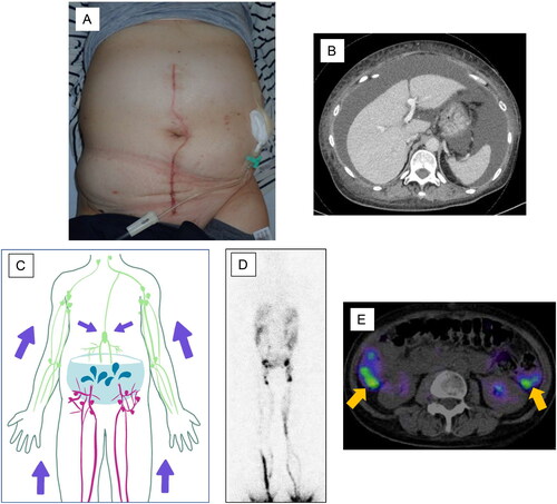
Figure 2. Intraoperative findings. Three lymphatic vessels and three veins are anastomosed in each of the right (A) and left (C) groin regions. The circles indicate the anastomoses. The triangle indicates the great saphenous vein (A). The tetragon indicates the accessory saphenous vein (C). The schema indicates the LVA on the right (B) and left (D) sides.
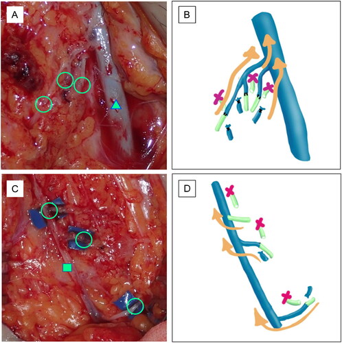
Figure 3. Postoperative findings. Abdominal distension disappears after LVA (A). Contrast-enhanced CT shows no evidence of ascites effusion (B). Lymphoscintigraphy (C) or SPECT (D) shows no evidence of radioisotope leakage into the peritoneal cavity. No secondary lymphedema is observed in either lower extremity. However, there is delayed lymphatic flow in the left lower extremity. The schema indicates that LVA reduced the amount of lymphatic fluid leaking into the abdominal cavity (E).