Figures & data
Figure 1. Left-hand and wrist radiography.
Complete amputation of left-hand digits one through four with a sustained an open fracture-dislocation of the fifth finger on that side. First and second metacarpal fractures, open proximal phalangeal fractures of the middle and ring finger, and open fracture dislocation through the DIP joint of the small finger were found. There was trans metacarpal thumb and index amputation and a cross-proximal phalanx of middle and ring finger amputation.
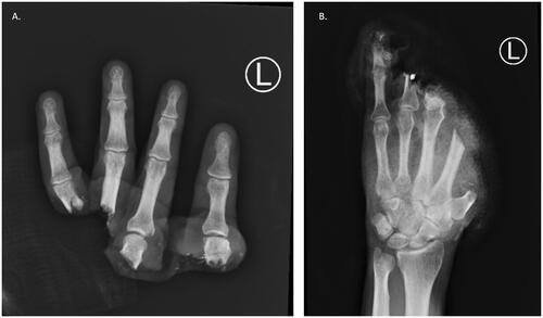
Figure 2. Amputated hand.
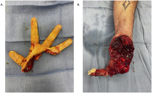
Figure 3. Vessels tagged in the amputated digits. (A) Radial digital artery to the thumb, radial artery in the snuffbox, ulnar digital artery to the index finger, radial digital artery to the middle finger, common digital artery to the third webspace, ulnar digital artery to the ring finger, radial digital artery to the fifth, and ulnar digital artery to the fifth. (B) Six anastomoses were performed into CV branches, using a vein coupler in a standard end-to-end fashion. For the thumb, in order to obtain adequate reach, two additional unrelated veins were utilized from the forearm.
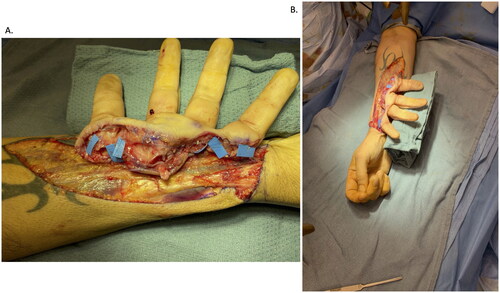
Figure 4. (A) 0.045-inch K-wires placed down each of the amputated digits in a retrograde fashion. (B) Osteosynthesis with K-wires was then performed, achieving bony rigidity and stability.
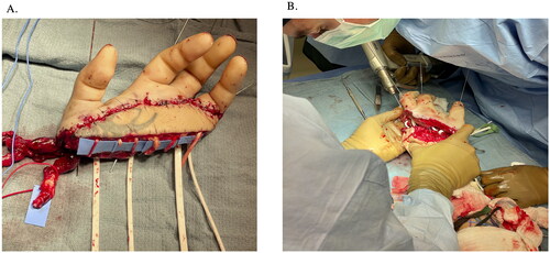
Figure 5. (A) 11-month follow-up in extension and (B) flexion.
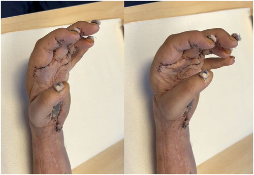
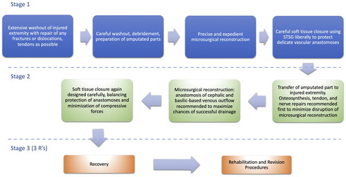
Figure 6. Flowchart outlining the main steps of temporary ectopic replantation in the upper extremity.