Figures & data
Figure 1. Dissection of the left wrist of an 85-year-old Korean female cadaver in the volar aspect. The anatomical structure of carpal tunnel was identified. We encountered an accessory first lumbrical muscle to the index finger within carpal tunnel (blue background). 1. Median nerve, 2. Accessory first lumbrical muscle, 3. Tendinous portion of the accessory first lumbrical muscle, 4. first lumbrical muscle, 5. Second lumbrical muscle, 6. Tendinous portion of flexor digitorum superficialis to the index finger.
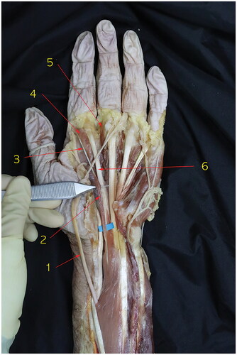
Figure 2. Dissection of the right wrist in the same cadaver with . 1. Tendinous portion of flexor digitorum superficialis to the index finger, 2. Origin of the accessory first lumbrical muscle in a tendinous form, 3. Accessory first lumbrical muscle bundle, 4. Tendinous portion of the accessory first lumbrical muscle, 5. first lumbrical muscle, 6. Second lumbrical muscle.
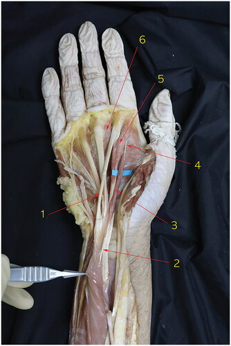
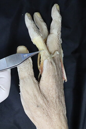
Figure 3. This reveals the insertion of the accessory first lumbrical muscle of the left index finger.
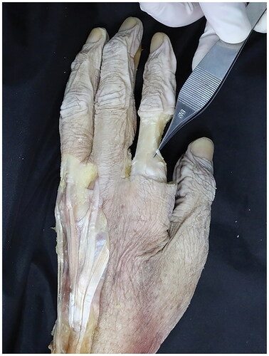
Figure 4. This reveals the insertion of the accessory first lumbrical muscle of the right index finger.