2,574
Views
23
CrossRef citations to date
0
Altmetric
Challenge Article

Temperature receptors in cutaneous nerve endings are thermostat molecules that induce thermoregulatory behaviors against thermal load

Pages 346-351 | Received 13 Feb 2015, Accepted 02 Apr 2015, Published online: 13 Oct 2015

Figures & data

Figure 1. Classical models of the thermoregulatory system. (A) The thermosensory system. It is assumed that thermoreceptor in cutaneous nerve ending is a thermosensor that sends nerve impulses as a temperature code to the brain, where the code is somehow decoded as “cold sensation.” (B) A thermostat in the brain. It is assumed that a thermostat (interneuron) exists in the brain based on the view that a skin thermoreceptor is a sensor to monitor skin temperature. This model states that the thermostat compares skin temperature with a set-point, and the error dependent output induces thermoregulatory responses. A circle with 2 inputs (+ and −) shows a comparator of the thermostat.
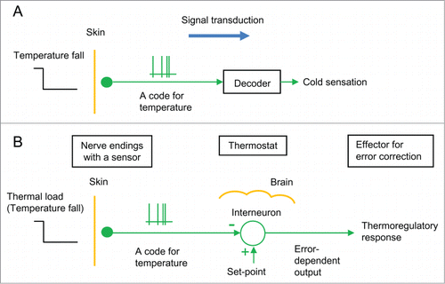
Figure 1. Classical models of the thermoregulatory system. (A) The thermosensory system. It is assumed that thermoreceptor in cutaneous nerve ending is a thermosensor that sends nerve impulses as a temperature code to the brain, where the code is somehow decoded as “cold sensation.” (B) A thermostat in the brain. It is assumed that a thermostat (interneuron) exists in the brain based on the view that a skin thermoreceptor is a sensor to monitor skin temperature. This model states that the thermostat compares skin temperature with a set-point, and the error dependent output induces thermoregulatory responses. A circle with 2 inputs (+ and −) shows a comparator of the thermostat.

Figure 2. A new model of a physiological thermostat. (A, B) A typical patch-clamp recording from sensory neurons responding to low temperatures (drawn referring to Okazawa et al.Citation16). (A) Whole-cell recordings. When the temperature falls below its whole-cell set-point (˜28 °C, threshold temperature), receptor potential occurs. When the receptor potential is above a threshold potential, action potentials occur. However, action potentials are inhibited when the receptor potential continues. Inset: time scale is expanded. (B) Single-channel recordings from an isolated patch membrane. When the temperature falls below its single-channel set-point (˜18°C, threshold temperature), single-channel activity appears.
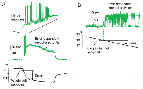
Figure 2. A new model of a physiological thermostat. (A, B) A typical patch-clamp recording from sensory neurons responding to low temperatures (drawn referring to Okazawa et al.Citation16). (A) Whole-cell recordings. When the temperature falls below its whole-cell set-point (˜28 °C, threshold temperature), receptor potential occurs. When the receptor potential is above a threshold potential, action potentials occur. However, action potentials are inhibited when the receptor potential continues. Inset: time scale is expanded. (B) Single-channel recordings from an isolated patch membrane. When the temperature falls below its single-channel set-point (˜18°C, threshold temperature), single-channel activity appears.

Figure 3. ThermoTRP channels as thermostat molecules. Error-dependent receptor potential of typical thermoTRP channels (Patapoutian et al.Citation11). The numbers in parentheses represent whole-cell set-points. TRPM8 and TRPA1 are thermostat molecules against low temperatures. TRPV1-4 are thermostat molecules against high temperatures.
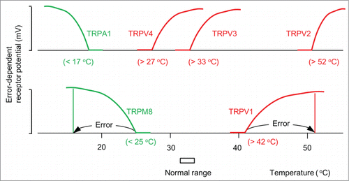
Figure 3. ThermoTRP channels as thermostat molecules. Error-dependent receptor potential of typical thermoTRP channels (Patapoutian et al.Citation11). The numbers in parentheses represent whole-cell set-points. TRPM8 and TRPA1 are thermostat molecules against low temperatures. TRPV1-4 are thermostat molecules against high temperatures.

Figure 4. A new model of the thermoregulatory system (Tajino et al.Citation17). Thermoregulatory system is composed of neural connection from a cutaneous nerve ending with thermostat molecules (TRPM8) to its target cerebral neurons for “cold” and heat-seeking behavior. A circle with 2 inputs (+ and −) shows a thermostat neuron. When skin temperature falls below a set-point, thermostat molecules in a nerve ending together generate error-dependent receptor potential for nerve impulses. These impulses run to the brain to activate its target neurons for “cold” and heat-seeking behaviors for error correction.
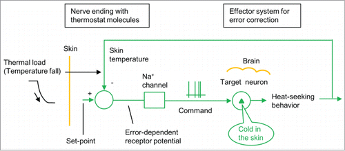
Figure 4. A new model of the thermoregulatory system (Tajino et al.Citation17). Thermoregulatory system is composed of neural connection from a cutaneous nerve ending with thermostat molecules (TRPM8) to its target cerebral neurons for “cold” and heat-seeking behavior. A circle with 2 inputs (+ and −) shows a thermostat neuron. When skin temperature falls below a set-point, thermostat molecules in a nerve ending together generate error-dependent receptor potential for nerve impulses. These impulses run to the brain to activate its target neurons for “cold” and heat-seeking behaviors for error correction.