Figures & data
Figure 1. Progenitor cell proliferation and migration in the hypothalamus Representative BrdU-labeled (red) sections of the hypothalamus inspected by laser-scanning confocal microscopy. HE6, HE13 and HE53 show samples on the 6th, 13th and 53rd day of heat exposure, respectively. 3V, third ventricle; scale bar, 50 µm. The photo samples are prepared using our previous data already published in a paper.Citation26
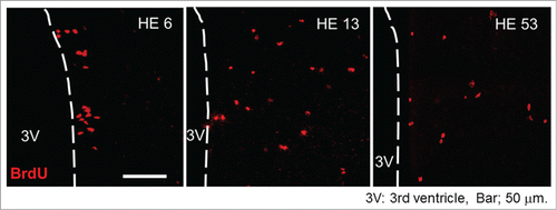
Figure 2. Newborn neurons in the hypothalamic area Representative BrdU (red) and NeuN (green) double-labeled sections of the hypothalamus inspected by laser-scanning confocal microscopy. HE43 and CN43 show samples on the 43rd day of heat exposure and of control, respectively. Yellow dots (shown as arrows) indicate BrdU and NeuN double-positive cells and therefore newborn neurons. 3V, third ventricle; scale bar, 100 µm. The photo samples are prepared using our previous data already published in a paper.Citation26
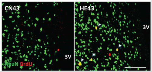
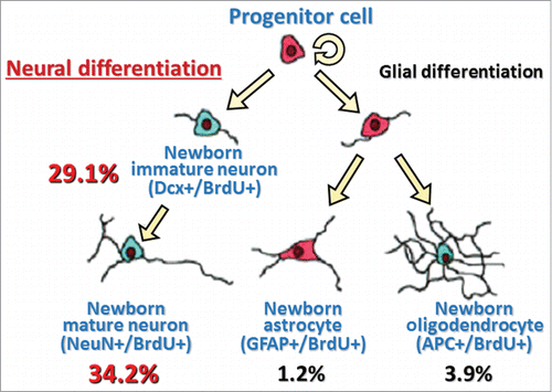
Figure 3. A model with summary for differentiation of heat exposure-induced newborn cells in the hypothalamus Values are percentages of respective differentiated cell number to total newborn cell number (values are obtained from our previous data already published in a paperCitation26). Dcx+/BrdU+, double-labeled with immature neuron marker (doublecortin, Dcx) and BrdU; NeuN+/BrdU+, double-labeled with mature neuron marker (neuronal N, NeuN) and BrdU; GFAP+/BrdU+, double-labeled with astrocyte marker (glial fibrillary acidic protein, GFAP) and BrdU; APC+/BrdU+, double-labeled with oligodendrocyte marker (adenomatosis polyposis coli, APC) and BrdU.