Figures & data
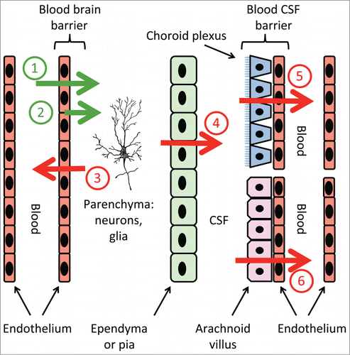
Figure 1. Transport pathways that might be involved when PGE2 signals fever in the brain. The green arrows show translocation of systemic PGE2 across the brain capillary endothelium, or the sided release of endothelium-derived PGE2, into the brain parenchyma. The red arrows show termination of PGE2 signaling via removal of PGE2. Numbers on arrows are discussed in the text.