Figures & data
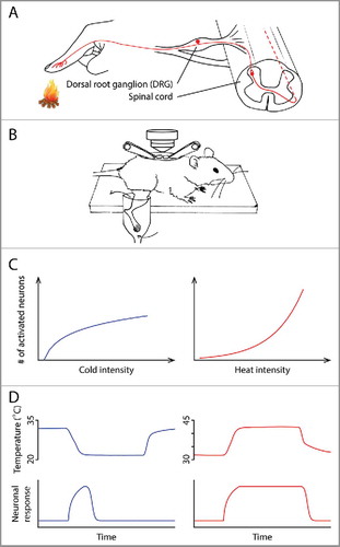
Figure 1. (A) Anatomy of the thermosensory pathway in the spinal cord and periphery. Ambient temperature is first detected by the free nerve endings of primary sensory neurons in the skin. These neurons, whose cell bodies are located in the dorsal root ganglia (DRG), relay information to the dorsal horn of the spinal cord, where thermal information is further processed before being sent to the brain. (B) A schematic illustrating the spinal cord imaging preparation, modified from CitationRef. 4. After a dorsal laminectomy, the lumbar vertebrae of an anesthetized mouse were stabilized using a pair of spinal clamps. The exposed spinal cord was loaded with a fluorescent calcium indicator, allowing in vivo optical recording of neuronal activity. The hind limb was superfused with water, the temperature of which could be precisely controlled. (C) The different temperature intensity-response relationships in the cold (left) and heat (right) ranges. (D) The different kinetics of cold and heat responses. Neuronal response to cold (bottom) correlates with the change of temperature (top), whereas response to heat correlates with the absolute temperature.