Figures & data
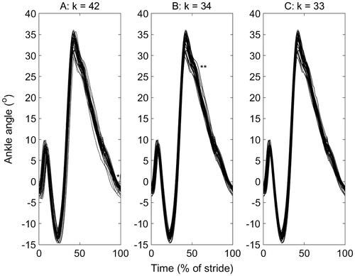
Figure 1. Ankle plantar-dorsi flexion angle for one participant on a standard treadmill running at 3.35 m/s: (a) before, (b), after stage 1 (b; α1 = 0.0001) and (c) after stage 2 (α2 = 0.01; b = 1) of a two-stage outlier removal method. The data were the right ankle from heel strike (0%) to heel strike (100%). The stance phase is approximately 0–40%. A potential spatial (*) and spatial–temporal (**) outliers are indicated, which are removed, and the number of cycles at each stage are indicated (k). The cycles that were deleted are listed here for stage 1 (3, 4, 5, 10, 26, 27, 41, 42) and stage 2 (11), which correspond to the column numbers in the supplementary material used to create this figure, to facilitate comparison with other outlier detection methods.