Figures & data
Figure 1. A. External and internal landmarks for electrode placement.
Notes: Cl: clavicle; Acr: acromion; IT: intramuscular tendon — approximate division of anterior and posterior regions of supraspinatus; ↔ muscle length. B. Electrode placement for anterior and posterior regions. * electrode placement for anterior region; X electrode placement for posterior region; 25% of muscle length from the medial aspect; 50% of muscle length.
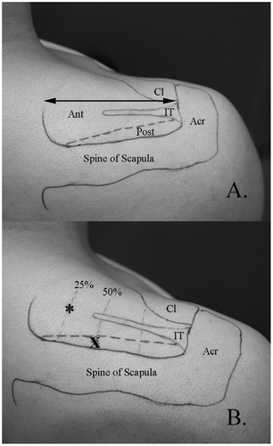
Figure 2. Fine-wire electrode placement.
Notes: Ultrasound guided insertion into the anterior region (A) and posterior region (B). Tp = trapezius; SP = supraspinatus; ***** intramuscular tendon; tip of needle.
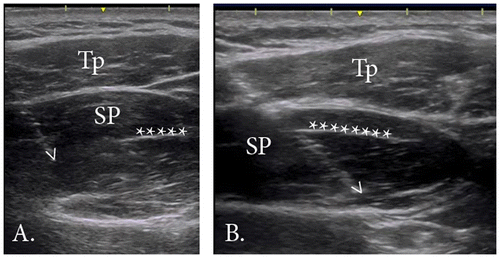
Figure 3. Summary of exertion and posture which demonstrated peak activity during MVIC for anterior and posterior regions. Abd = humeral abduction; ER = external rotation.
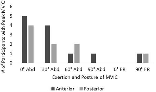
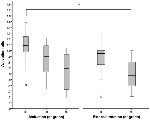
Figure 4. Median ratio (anterior: posterior) for abduction and external rotation exertions in various postures. Significant difference (p < 0.005). Outliers indicated by o.