Figures & data
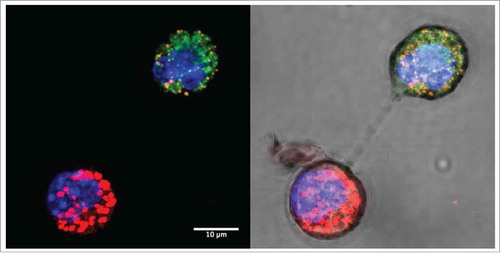
Figure 1. Intercellular mitochondrial transfer. Confocal images of mitochondrial transfer between MitoTracker-labeled 4T1 breast carcinoma cells (red) and 4T1ρ° cells (green) under dark field (left) and bright field (right) showing membrane nanotube connections. Cells were co-cultured for 24 h and nuclei were stained with NucBlue.