Figures & data
Figure 1. The Med1 expression was regulated through DUBs overexpression.
To determine Med1 ubiquitination, the total 293T cell lysates were performed immunoprecipitation (IP) with anti-Med1 antibody or anti-IgG as negative control, and immunoblotting with anti-Ub antibody and anti-Med1 (A). Several DUB DNA plasmids were transfected into MCF7 breast cancer cell. The Med1 protein expression was confirmed by western blotting using anti-Med1 and anti-SRT to compare the level of control vector transfected cells and normalized with MCF7 control cells (B). The transcriptional activity of Med1 was measured by ERE-Luc reporter luciferase plasmid (C). All experiments were repeated three times, and the presented data are the mean ± standard error of the mean (SEM). * p < .05, ** p < .01, compared with MCF7 control cells.
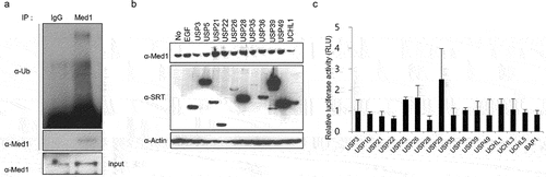
Figure 2. Med1 protein and mRNA expression through BAP1 overexpression.
To determine Med1 protein expression, MCF7 cells were transfected with SRT-USP21, SRT-USP35, SRT-BAP1 wild-type (WT) or SRT-BAP1 C91A mutant-type (MT) DNA plasmids. The total cell lysates were immunoblotted with the anti-Med1 and anti-SRT. Expression of β-actin was used as a loading control. (A, B) Med1 mRNA expression was performed by real-time PCR. The Med1 mRNA expression was normalized with control vector-introduced cells (C). The MCF7 cells were harvested for 24 h after treatment with 10 ng EGF, E2, TGF-β, and then the total cell lysates were immunoblotted with anti-Med1 and anti-BAP1 (D). The values shown are the means of three independent experiments. The presented data are the means ± standard error of mean (SEM). *p < .05, **P < .01 compared with MCF7 control cells.
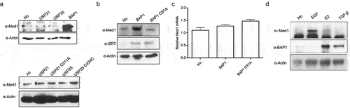
Figure 3. Med1 interacted with BAP1 and regulated growth and metastatic ability.
To determine Med1 and BAP1 interaction, 293T cells were transfected with BAP1 or BAP1 C91A DNA plasmids for 36 h and then the cell lysates were immunoprecipitated with anti-Med1 antibody and subsequently immunoblotted with anti-SRT antibody or immunoblotted with anti-BAP1 (A). Cell proliferation was measured by counting the number of trypan-blue staining cells at 3 d and 6 d after transfecting SRT-BAP1 or SRT-BAP1 C91A DNA plasmids into MCF7 cells. All proliferation assays were repeated three times (B). The Matrigel cell invasion assay was performed using SRT-BAP1 WT or SRT-BAP1 C91A DNA plasmids and compared with MCF7 control cells. After incubating the cell with Matrigel-coated filters for 24 h, the cells on the undersurface of the filter were staining with hematoxylin and eosin (H&E stain). The invaded cell measured OD values using spectrophotometer. The cell was starved in serum-free media for 24 h before being to the manufacturer’s instructions. The migrated cells were stained, and OD values were measured using spectrophotometer. Quantitative RT-PCR (qPCR) analysis was performed using gene-specific primer in BAP1 or BAP1 C91A DNA plasmids transfected cells to confirm the observed alteration of uPAR target gene expression. Later, the mRNA level of each gene was normalized to the level of β-actin mRNA. The relative mRNA was compared with MCF7 control cells (C).
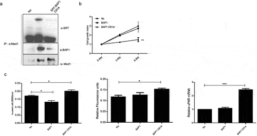
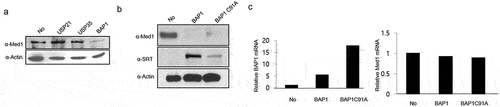
Figure 4. Med1 protein and mRNA expression through BAP1 overexpression in lung cancer cells.
To determine Med1 protein, A549 cells were transfected with SRT-USP21, SRT-USP35, SRT-BAP1 wild type (WT) or SRT-BAP1 C91A mutant type (MT) DNA plasmids. The total cell lysates were immunoblotted with the anti-Med1 and anti-SRT. Expression of β-actin was used as a loading control (A, B). Med1 and BAP1 mRNA expressions were performed by real-time PCR. Expression of β-actin was used as a loading control. The Med1 and BAP1 mRNA expressions were normalized with control vector introduced cells (C).