Figures & data
Fig. 1 Discrete wavelet transform with three levels.
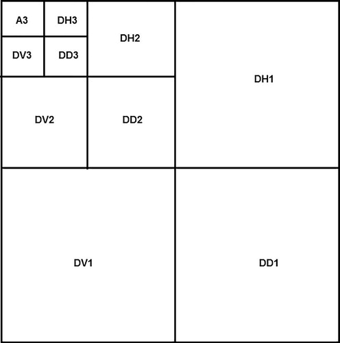
Fig. 2 A slice of simulated human brain with resolution 128 × 128.
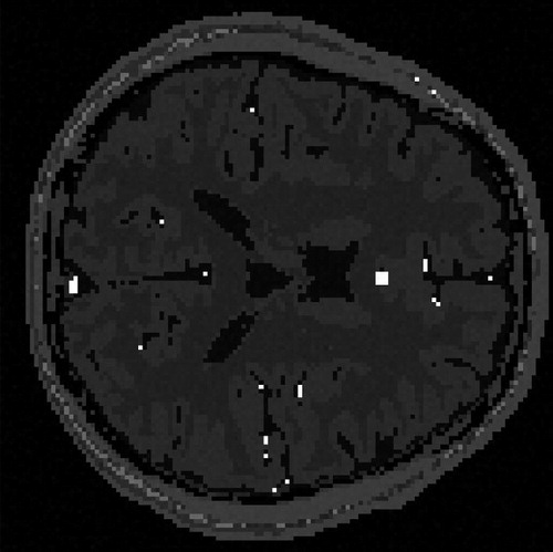
Fig. 3 Wavelet transformed image. Left: Discrete wavelet transformed image. Right: The sparsified image.
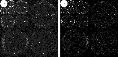
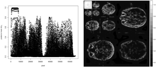
Fig. 4 Posterior mean of pi for every pixel Left: Scatter plot of the posterior mean of pi. Right: Image form of the posterior mean of pi.
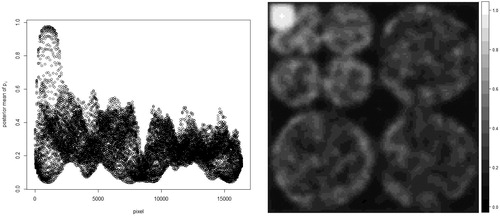
Fig. 5 for 90 simulations.
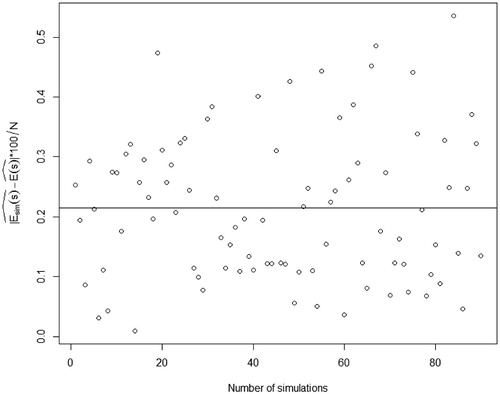
Fig. 6 Normal quantile-quantile plot of standardized from 90 simulations.
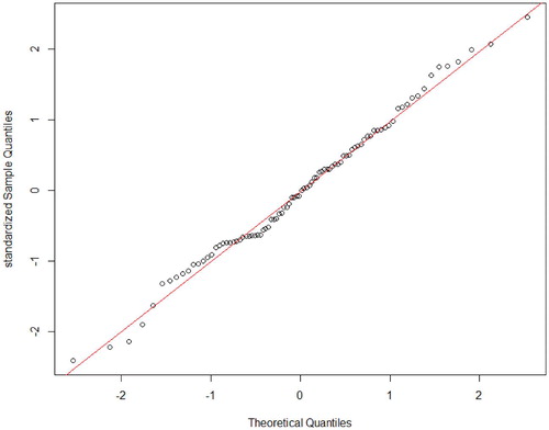
Fig. 7 One slice of real human brain with resolution 256 × 256.
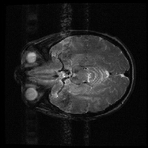
Fig. 8 Wavelet transformed image Left: Discrete wavelet transformed image. Right: The sparsified image.
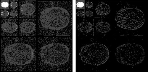
Fig. 9 Posterior mean of pi for every pixel. Left: Scatter plot of the posterior mean of pi. Right: Image form of the posterior mean of pi.