Figures & data
Figure 1–4. Macugonalia semiguttata (Signoret, 1853). 1–3, body, in dorsal and lateral view, and face, anterior view (male). 4, original illustration provided by Signoret (1853, pl. 12, Figure 4) showing diagnostic color features of the species.
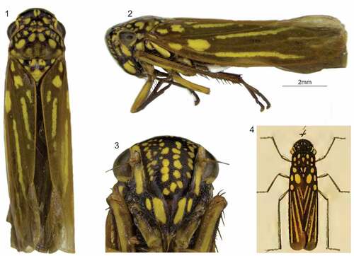
Figure 5. Macugonalia semiguttata (Signoret, 1853), specimen photographed on a shrub beside the road to the Pico da Caledônia, Nova Friburgo, Rio de Janeiro State (collection site is 1,737 m a.s.l.); photograph taken by André Almeida Alves, 6 December 2019.
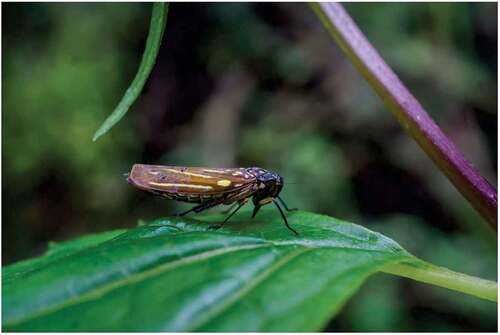
Figure 6–9. Macugonalia semiguttata (Signoret, 1853), male. 6, apical portion of abdomen, ventral view. 7, pygofer, lateral view. 8, subgenital plates, connective, and styles, dorsal view. 9, aedeagus, lateral view.
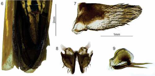
Figure 10–14. Macugonalia semiguttata (Signoret, 1853), female. 10, apical portion of abdomen, lateral view (the sternite VII is dislocated downward and the ovipositor is exposed). 11, sternite VII, ventral view. 12, valvifer I and valvula I, lateral view; 12a, dorsal sculptured area at apical portion; 12b, apex. 13, valvula II, lateral view; 13a–b, teeth at median and apical portions; 13 c, apex. 14, valvifer II and gonoplac, lateral view. DEN: denticle; DSA: dorsal sculptured area; DUC: duct; Evii: sternite VII; GON: gonoplac; OVI: ovipositor valvulae I and II; RAM: ramus; TOO: tooth; VID: ventral interlocking device; Vli: valvifer I; Vlii: valvifer II; VSA: ventral sculptured area.

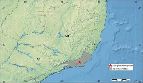
Figure 15. Collection site of Macugonalia semiguttata (Signoret, 1853) in the Serra dos Órgãos mountain range, Rio de Janeiro State (22°20ʹ44.8”S, 42°35ʹ13.2”W, circa 1,700 m a.s.l.). This is the single known precise record of the species. ES: Espírito Santo State; MG: Minas Gerais State; SP: São Paulo State.