Figures & data
Figure 1. Localities sampled where species of microturbellarians are found in Peru. A) Species included in this study, B) species from du Bois-Reymond Marcus [Citation28], C) species from Reyes & Brusa [Citation5], D) species from Reyes et al. [Citation30], E) species from Noreña et al. [Citation29,Citation34] and Damborenea et al. [Citation22], F) species from Damborenea et al. [Citation24], and G) species from Beauchamp [Citation27].
![Figure 1. Localities sampled where species of microturbellarians are found in Peru. A) Species included in this study, B) species from du Bois-Reymond Marcus [Citation28], C) species from Reyes & Brusa [Citation5], D) species from Reyes et al. [Citation30], E) species from Noreña et al. [Citation29,Citation34] and Damborenea et al. [Citation22], F) species from Damborenea et al. [Citation24], and G) species from Beauchamp [Citation27].](/cms/asset/af109cfc-96ba-4bd3-b1cb-0fdb17138a37/tneo_a_2040348_f0001_oc.jpg)
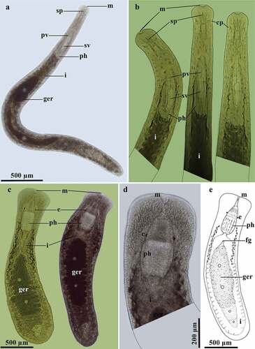
Figure 2. Stenostomum grande Child, 1902. A, Photograph of a live specimen; B, Schematic representation of the habitus; C, Detail of the anterior region of the body; D, Detail of the region of the pharynx; E, Detail of the caudal region of the body.
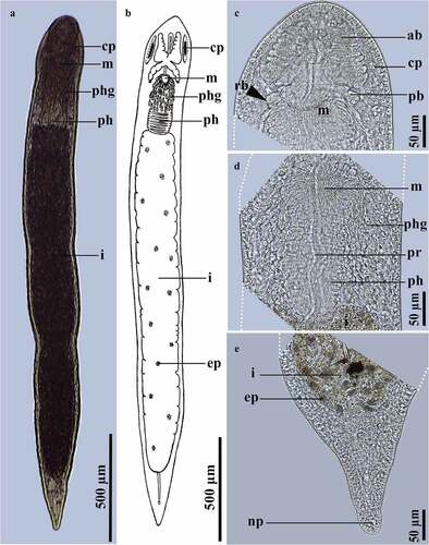
Figure 3. Photographs of live species of Stenostomidae in ventral view. A, Stenostomum saliens Kepner & Carter, 1931; B, S. tuberculosum Nuttycombe & Waters, 1938 with detail of the posterior end of the body covered by cilia (inside the square); C, Myostenostomum vanderlandi Rogozin, 1992.
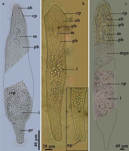
Figure 4. Gieysztoria cuspidata (Schmidt, 1861) Ruebush & Hayes, 1939. A, Dorsal view of a squashed live animal showing its internal organs; B, Schematic representation of the habitus of a live swimming animal showing dorsal pigment; C, Penis stylet from a whole-mounted individual (yellow arrowheads indicate each spine); D, Schematic representation of the penis stylet with detail of the digitiform projections.

Figure 5. Gieysztoria bellis (Marcus, 1945). A, Dorsal view of a squashed live animal; B (1–4), Penis stylet of a whole-mounted individual in different focal planes with some bent spines and weak distal rings (same scale bar for all photographs); C, Detail of the male reproductive system of a whole-mounted individual; D (1–2), Penis stylet of a whole-mounted individual in different focal planes; E, Schematic representation of the penis stylet in photograph B (1–4); F, Schematic representation of the penis stylet in photograph D (1–2).
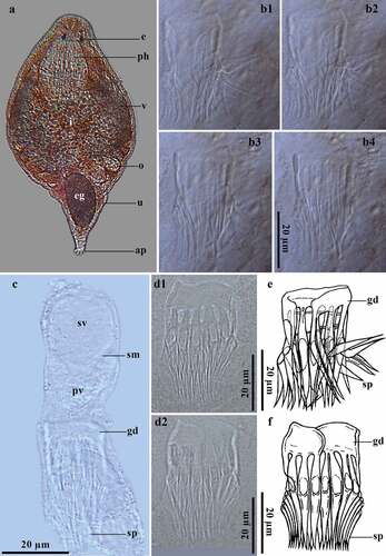
Figure 6. Prorhynchus stagnalis Schultze, 1851. A, Dorsal view of the habitus; B, Detail of the anterior end of the body in sequence of movements (no scale bar available). Geocentrophora applanata (Kennel, 1888). C, Dorsal view of the habitus, sequence of movement; D, Detail of the anterior region of the body; E, Schematic representation of the habitus.