Figures & data
Figure 1. Axial CT showed a mucocele on the center of the frontal lesion, thinning the rear bony wall of the frontal table in the midline (A). MRI showed that the frontal mass had heterogeneous moderate to high intensity on T1-weighted and moderate to low intensity on T2-weighted imaging (B, C). The remnants of the bilateral small frontal sinuses were located on the both sides of the cyst, on the right side with air (A, D: white arrows) and on the left side with soft tissue (E: black arrow).
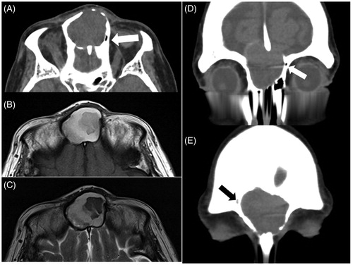
Figure 2. CT demonstrated that the lesion existed in both of the frontal sinuses, invading the left orbit by destroying the orbital roof (A), and there was a ‘niche’ at the center of the frontal skull base instead of the frontal septum (A: black arrow). The frontal sinus septum was mostly destroyed but partially remained in a ‘Y’ shape, which implied ballooning and bursting of the mucocele of ISSC into both sides of the frontal sinus (B, C: black arrow). MRI showed a moderate-intensity mass in the middle of the frontal lesion surrounded by a high-intensity area on T1-weighted imaging (D).
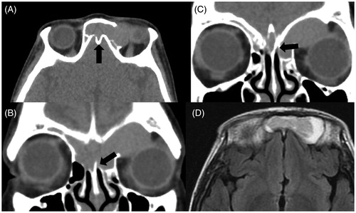
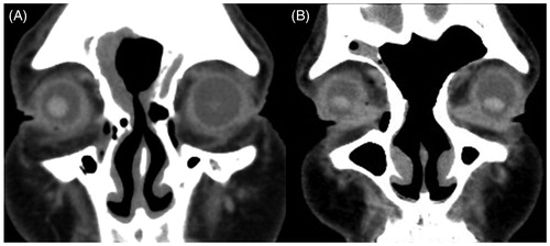
Figure 3. Postoperative CT scans of case 1 (A) and case 2 (B). The outflow tracts were maintained in both cases.