Figures & data
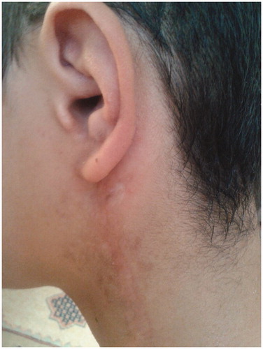
Figure 1. (A, B) A neurofibroma filling the external ear canal and pushing the auricle forward. C: Coffee-coloured stains on the skin.
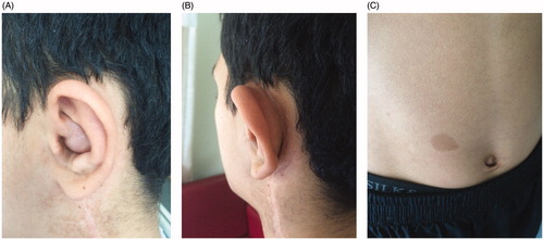
Figure 2. Preoperative audiogram of the patient.
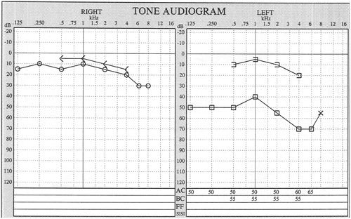
Figure 3. (A) On axial temporal bone CT, the right external ear canal is open. In the left ear, a soft tissue mass (white arrows) surrounds and obliterates the external ear canal between the region of the left temporal bone mastoid (black arrow) and the mandible (white arrowhead). (B) On the more inferior slices, the mass approximates the internal carotid artery (red circle), the internal jugular vein (blue circle), and the superior parapharyngeal area (star). (C) The mass approaches the annulus to within 3 mm (black arrow) and obliterates the external ear canal.
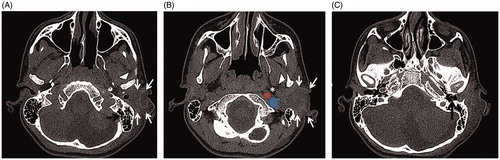
Figure 4. (A, B) A neurofibroma measuring approximately 5 × 4 × 3 cm, exhibited bleeding, lacked a capsule, and showed dense infiltration of the surrounding tissue. The neurofibroma was typical in appearance, resembling a ‘bag of worms’. (C) The external auditory canal after surgery.
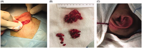
Figure 5. A T1-weighted magnetic resonance image obtained 2.5 years postoperatively shows that both external ear canals are open and the surrounding soft tissues are normal.
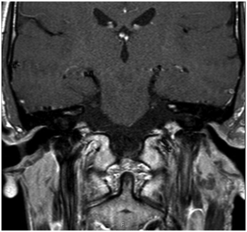
Figure 6. The auricle and external auditory canal photographed 2 years postoperatively.