Figures & data
Figure 1. Pure-tone audiogram. His right hearing loss was progressed in one month. O: right air-conducted hearing level; X: left air-conducted hearing level; [: right bone-conducted hearing level; ]: left bone conducted hearing level.
![Figure 1. Pure-tone audiogram. His right hearing loss was progressed in one month. O: right air-conducted hearing level; X: left air-conducted hearing level; [: right bone-conducted hearing level; ]: left bone conducted hearing level.](/cms/asset/291072db-434f-454f-8c06-2f040d553748/icro_a_1319735_f0001_c.jpg)
Figure 2. VEMP findings. 500 Hz short tone bursts (air-conducted, 125 dBSPL). cVEMPs were recorded on the sternocleidomastoid muscle ipsilateral to the stimulated ear, while oVEMPs were recorded just below the lower eye lid contralateral to the stimulated ear. Both of cVEMP and oVEMP were absent to the right ear stimulation (air-conducted 500 Hz short tone bursts, 125 dBSPL).
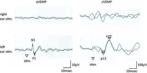
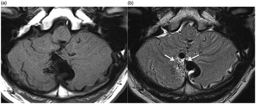
Figure 3. MRI findings. a. T1 weighted imaging. b. Flair imaging. Arrows indicate the flow void of AVM nidus located in the cerebellar vermis.