
Figures & data
Figure 1. ANP presenting as a large exophytic mass, left nasal polyp extending posteriorly into the oropharynx too. *Picture used after patient and relatives consent.
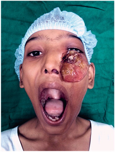
Figure 2. Pre-operative CT PNS.
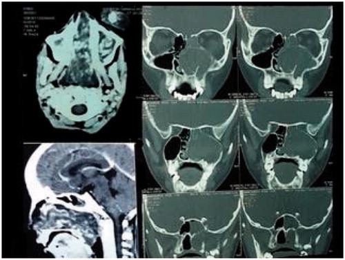
Figure 3. Excision biopsy of the exophytic mass.
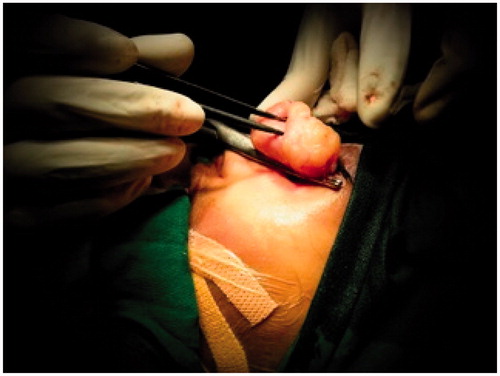
Figure 4. Complete excision and clearance of the sinuses in piece-meal fashion.
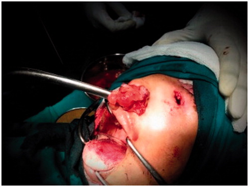
Figure 5. Excised polypoidal mass.
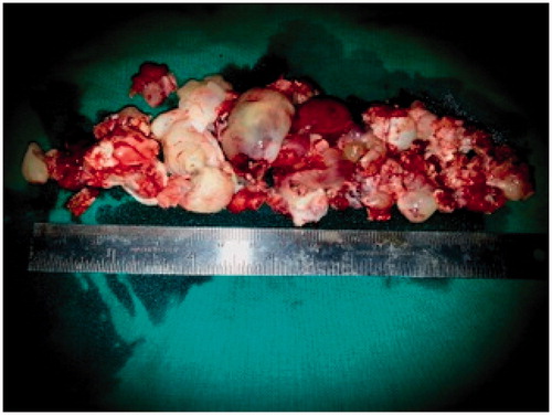
Figure 6. Suturing of the external skin defect and nasal packing over the balloon of Foley’s catheter.
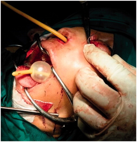
Figure 7. Histopathology showing atypical spindle cells and ectatic blood vessels in eosinophilic stroma.
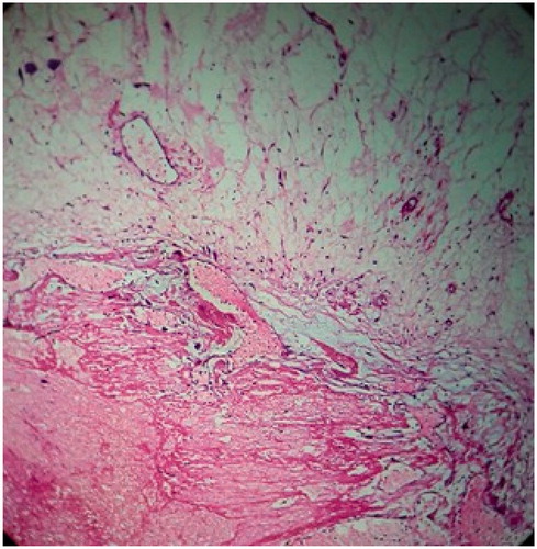
Figure 8. Post-operative CT PNS after 4 weeks.
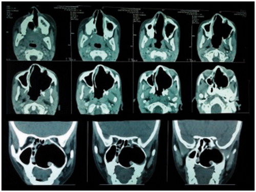
Figure 9. Post-operative scar and zero degree endoscopic view of the left nasal cavity, after 4 weeks. *Picture used after patient and relatives consent.
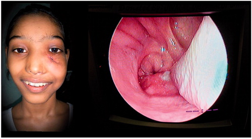
Table 1. Differential diagnosis of angiectatic sinonasal polyps.