
Figures & data
Figure 1. (Mid) sagittal section of CT-scan of the head and neck area revealing a foreign body (arrow) of ±5 cm in length and 1.5 cm across at the level of the hypopharynx and the proximal oesophagus.
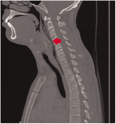
Figure 2. Direct laryngoscopy shows a fin-like structure lateral of the left arytenoid with supraglottic oedema. Intubation with a single-lumen tube.
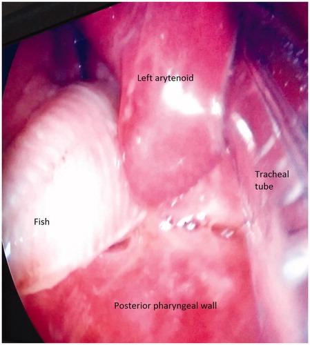
Figure 3. Sagittal CT slice shows a residual foreign body (arrow) at the posterior wall of hypopharynx.

Figure 4. Remaining pectoral fin in the left hypopharynx.
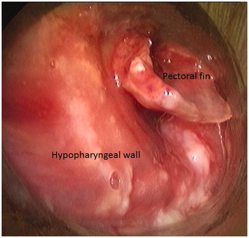
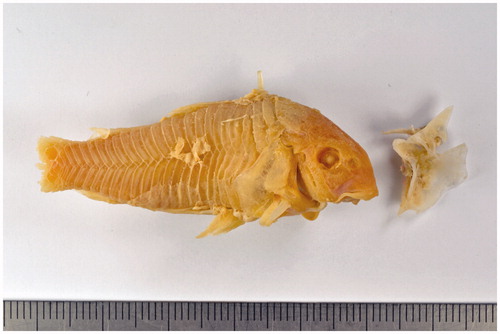
Figure 5. Bronze catfish (Corydoras aeneus), lacking the tail (left) and pectoral fin (right) as recovered from the patient; preserved in the collection of the Natural History Museum Rotterdam.
Supplemental material