Figures & data
Figure 1. Computed tomography shows a homogenous and well circumscribed soft tissue mass in the left external auditory canal (white arrows).
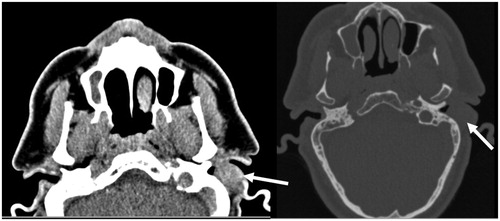
Figure 2. Surgical exploration showed a well circumscribed, not encapsulated soft tissue mass of 2 × 2 × 1 cms.
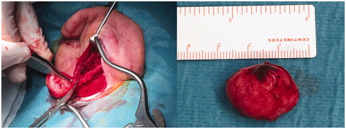
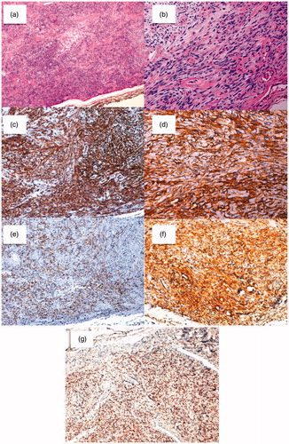
Figure 3. Histological sections. (3a): Patternless pattern of hyper and hypocelullar areas separated by collagen bands (Hematoxylin and Eosin (H&E) magnification 10×); (3b): hypercelullar area with spindle-like cells (H&E magnification 20×). On immunohistochemical analysis; (3c): tumor cells were highly reactive to CD34 (magnification 10×); (3d): CD99 (magnification 20×); (3e): Bcl-2 (magnification 10×); (3f): vimentin (magnification 10×); (3g): Positive STAT6 nuclear expression (magnification 10×).