Figures & data
Figure 1. Tumors of the external auditory canal and auricle. (a, b)At the initial visit, a painless mass with a smooth surface was found in the right external auditory canal which was almost completely occluded. Also, several tender masses with severe pain were observed around the right auricle. (a) Inside the right ear. (b) Behind the right ear. (c, d) Three years after surgery, no recurrence is observed either in the ear or the subcutaneous region behind the ear. (c) Inside the right ear (after surgery). (d) Behind the right ear (after surgery).
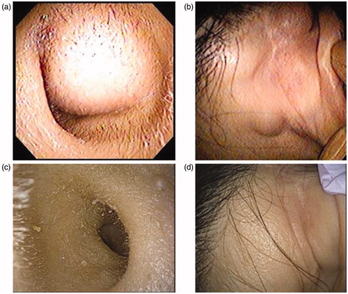
Figure 2. Pure tone audiometry. (a) Before surgery, conductive hearing loss of the right ear was observed. (b) After surgery, the right air-bone gap completely disappeared.
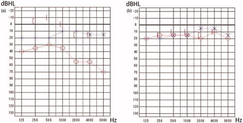
Figure 3. Preoperative images. (a, b, c, d) In CT, a 2 cm tumor on the anterior wall of the right external auditory canal with the destruction of the bones of the anterior and inferior walls was found. Also, multiple masses were observed behind the right ear. (e, f) In contrast-enhanced MRI, a tumorous lesion with a diameter of 19 mm in the right external ear canal, as well as multiple well-defined masses above, behind, and below the right auricle were found. Also, a suspected small vestibular schwannoma localized in the left internal ear canal was observed.
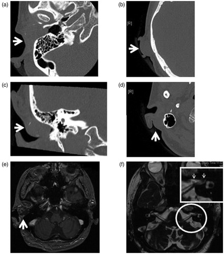
Table 1. Diagnostic criteria of Schwannomatosis.
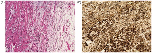
Figure 4. Histopathological examination. (a) Compact spindle cells arranged in short bundles, composed of focal hypercellular (Antoni-A) areas and hypocellular areas (Antoni-B) with no malignant findings (H–E). (b) The tumor cells are positive for S-100 (Immunohistochemistry). Scale bar: 100 μm in (a) and (b).