Figures & data
Figure 1. Initial axial CT scans without contrast of paranasal sinus. Although CT showed mild mucosal swelling or partial opacifications in bilateral sinuses, it was hard to identify the causative legion of optic neuropathy.
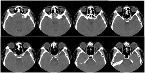
Figure 2. (a) T1-weighted, (b) T2-weighted and (c) Gd-enhanced coronal MRI images of paranasal sinus. MRI revealed a cystic lesion (arrow) around left anterior clinoid process which was adjacent to the left optic canal. There was an arachnoid cyst in the middle cranial fossa (arrowhead).
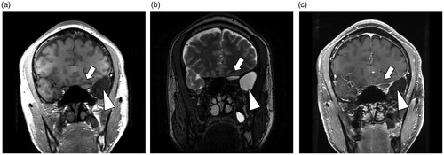
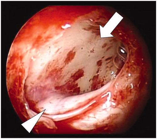
Figure 3. Intraoperative view of the cyst (arrow) and left optic nerve canal (arrowhead), both of which were opened endoscopically.