Figures & data
Figure 1. Our bucket used for the “bucket test.”
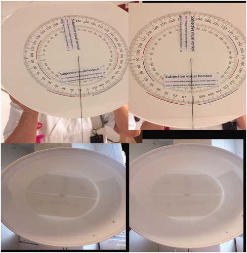
Figure 2. Coronal CT angiography and 3D reconstruction: symmetric opacification of AICA. No occlusion is shown.
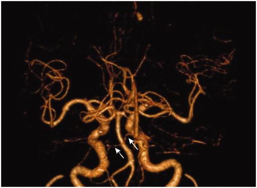
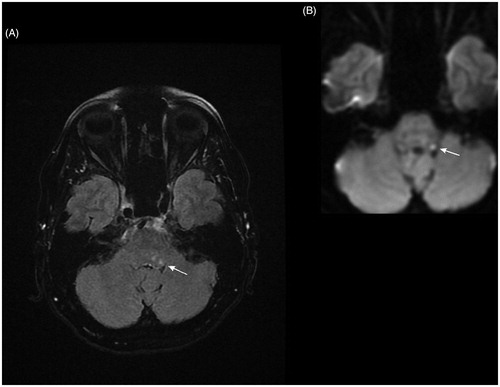
Figure 3. (A) MRI FLAIR (fluid attenuation inversion recovery) and (B) DWI (diffusion weighted imaging) axial slices show a small focus of high signal in the left facial colliculus, suggesting acute ischemia.