Figures & data
Figure 1. Case 1. (A) Preoperative CT scan—axial cut—demonstrating extensive soft tissue gas involving the neck and chest wall. (B) Preoperative examination with erythema and swelling involving the neck and chest. (C) Preoperative airway evaluation with edema and airway compromise. (D) Patient after first debridement and tracheostomy.
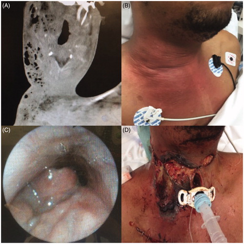
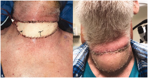
Figure 2. Case 1. (A) Extensive thoraco-cervico-facial soft tissue defect and great vessel exposure following multiple debridements. (B) Immediate postoperative appearance following reconstruction with an anterolateral thigh free flap. (C) Patient appearance at follow-up.
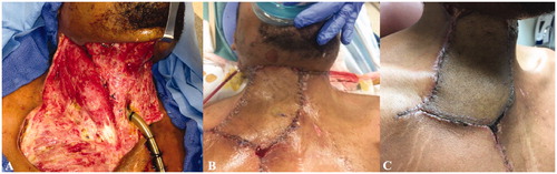
Figure 3. Case 2. (A) Pre-operative CT scan—axial cut—demonstrating extensive soft tissue gas involving the bilateral neck with extension into the anterior mediastinum. (B) Extensive soft tissue defect involving bilateral neck with exposure of larynx and great vessels.
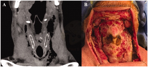
Figure 4. Case 2. Reconstruction with an anterolateral thigh free flap. (A) Immediate postoperatively and (B) at follow-up.