
Figures & data
Figure 1. Contrast-enhanced CT findings at first visit and MRI findings 3 months after first visit. Contrast-enhanced CT at first visit showed one tumor in the upper end (A, arrowhead) and two tumors in the lower end (B, arrowhead) of parotid gland. MRI findings 3 months after the first visit showed enlarged tumors in axial section of T1 weighted intensity (C,D) and coronal section of T2 weighted intensity (E,F). Arrowheads indicate the tumors in the upper end (C,E) and the lower end (D,F) of parotid gland and superior deep lateral cervical lymph node (F).
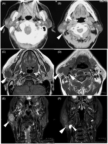
Figure 2. Intraoperative finding and resected specimen. Arrowheads indicate the main trunk of the facial nerve at the cross point (A). The upper end of tumor (white dotted circle) invaded the zygomatic branch of facial nerve. The surgical specimens were whitish in color (B).
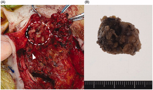
Figure 3. Microscopic finding of resected tumor. (A) Hematoxylin and Eosin (HE) stained finding in low power field. (B) HE stained finding in high power field. (C) S-100 immunostained finding. (D) Langerin immunostained finding. (E) CD21 immunostained finding.
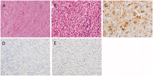
Table 1. Reported cases of IDCS of the parotid gland.