Figures & data
Figure 1. Contrast-enhanced computed tomography scan showing remnants of glass pieces in the neck (arrow) and the damaged right vertebral artery and pseudoaneurysm (arrow head).
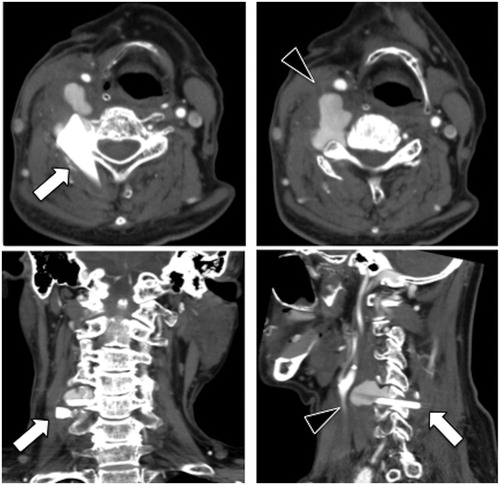
Figure 2. (A) The vertebral artery pseudoaneurysm (arrow head). (B) Successful obstruction of the right vertebral artery using coil embolization.
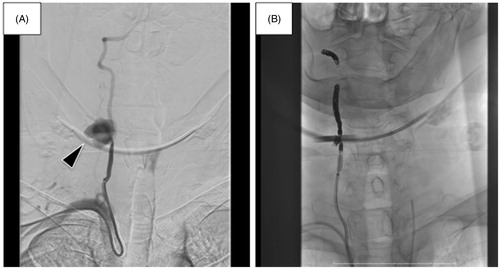
Figure 3. (A) The right neck exploration. The arrow head shows the edge of the glass. (B) The retrieved glass pieces. The larger one was located between the transverse processes of C4 and C5.
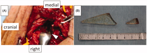
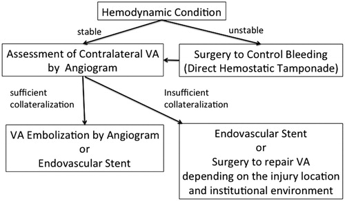
Figure 4. Flow chart for treatment decision-making.