Figures & data
Figure 1. Endoscopic-assisted arytenoid adduction surgery (EAAS). From the cricothyroid ligament toward the piriform sinus, the penetration needle and the loop needle are inserted, and the retracted nylon threads are tightened with a spacer at the cricothyroid ligament. The arytenoid cartilage is successfully adducted. This figure is modified from Murata et al. [Citation7].
![Figure 1. Endoscopic-assisted arytenoid adduction surgery (EAAS). From the cricothyroid ligament toward the piriform sinus, the penetration needle and the loop needle are inserted, and the retracted nylon threads are tightened with a spacer at the cricothyroid ligament. The arytenoid cartilage is successfully adducted. This figure is modified from Murata et al. [Citation7].](/cms/asset/ac0f36f0-fa37-43ca-8065-0efd1da6968f/icro_a_1655429_f0001_b.jpg)
Figure 2. Schema of the fenestration approach. Arytenoid adduction (AA) is performed through the fenestration on the thyroid ala. The suture is pulled in the direction of lateral cricoarytenoid muscle (LCA) contraction and fixed to the cricoid cartilage.
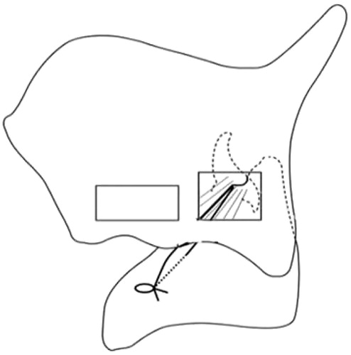
Table 1. Pre- and post-operative results.
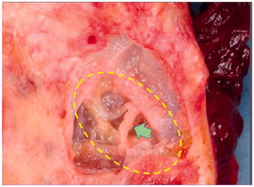
Figure 3. Blood vessel running outside the arytenoid cartilage. Care is taken to avoid damaging blood vessels outside the lateral cricoarytenoid muscle accompanying the adduction branch of the recurrent laryngeal nerve.