Figures & data
Figure 1. The facial nerve occupied the atresia plate and part of the promontory (a, e) with dehiscence. The titanium stapes prothesss was shaped with two curves in different planes (b, f). The prothesis connected the scale tympani opening and the manubrium of malleus (c, f). The connection of the chain with 45° hook (d). I: incus; M: malleus; P: prothesis; C: cochleostomy; FN: facial nerve; RW: round window; TM: tympanic membrane. Black arrow: cochleostomy on the lateral wall of basal scala tympani.
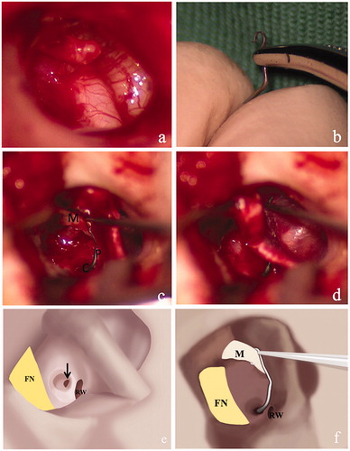
Figure 2. Cone-bean CT at 1 month after surgery. Axial section showing the absence of oval window (black arrow) with the facial nerve occupying the vestibular area in the affected side (a) vs. the normal oval window in the unaffected side (black arrow) (b). The stapes prothesis was inserted into the scala tympani along the superior margin of the round window (c) and the white arrow showed the round window niche. From the coronal image section, the length of protrusion was scaled (d).
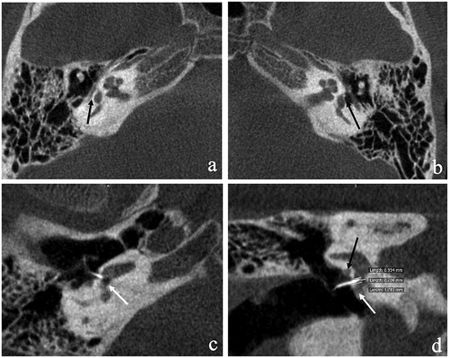
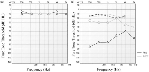
Figure 3. Audiogram at 3 months after surgery. Unaffected side (a). Hearing improvement in the affected side (b). black lines: prior to surgery. Grey lines: after surgery.