Figures & data
Figure 1. (A) Pure tone audiogram of the patient after the first septic meningitis. (B) Preoperative pure tone audiogram of the patient, having suffered second sepsis.
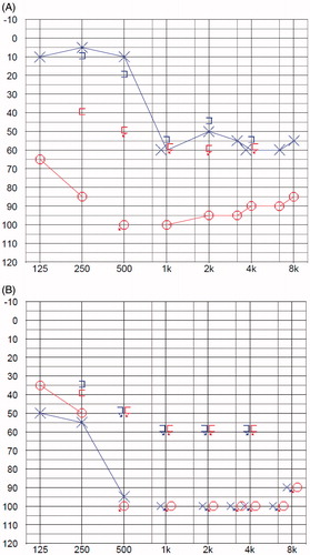
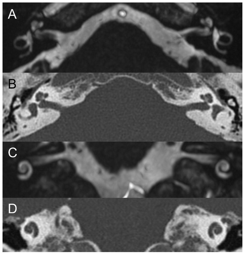
Figure 2. Preoperative MRI (A, axial; C, coronal) and CT (B, axial; D, coronal) showed normal cochlear and vestibular fluid spaces, with no imaging evidence of labyrinthitis. Specifically, MRI showed no abnormal obliteration, signal change, or contrast enhancement of fluid spaces, and CT did not show ossification.