Figures & data
Figure 1. Preoperative images. (A) Preoperative picture, the with arrow points at the sinus of the cartilaginous auditory canal. (B) Preoperative otoscopic image.
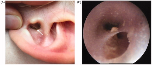
Figure 2. Preoperative CT-scan. Consecutive axial sections, the white arrow points at the sinus of the cartilaginous auditory canal.
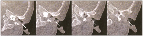
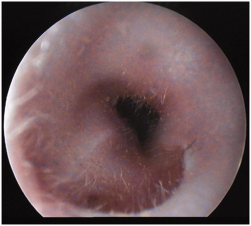
Figure 3. Postoperative otoscopic image. This otoscopic image was taken at 3 months follow-up.