Figures & data
Figure 1. A nasal endoscopy shows a bulge from the left floor of the nose to the bottom of the nasal septum, but there is no bulge in the palate.
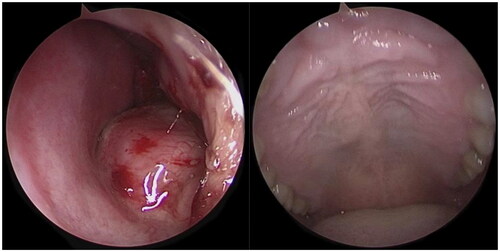
Figure 2. CT shows round-like swelling changes from the palatine bone to the base of the nasal septum.
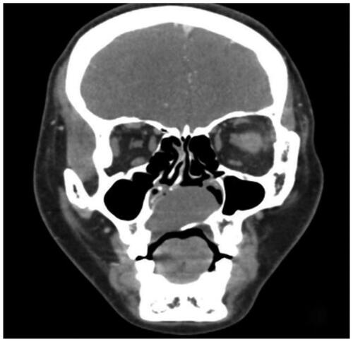
Figure 3. MRI shows a cyst in the palate.
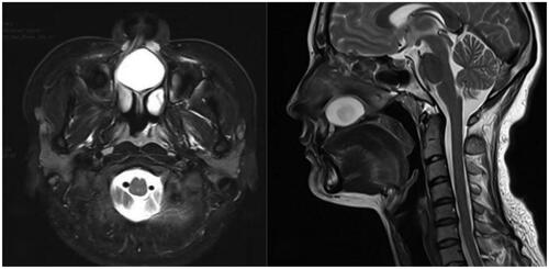
Figure 4. The nasal endoscopic incision of the nasal fundus mucosa shows cholesterol crystals.
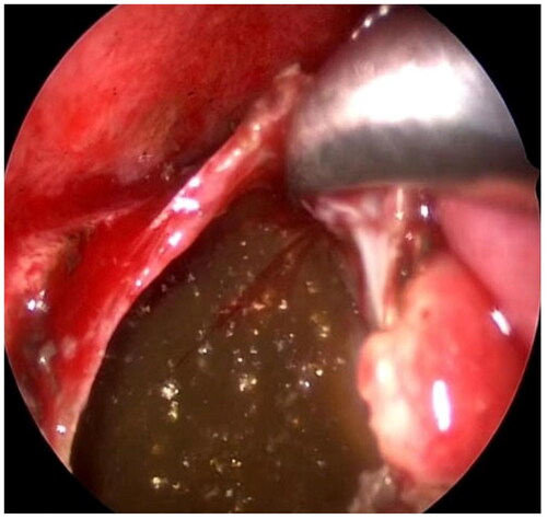
Figure 5. Both nasal floor flaps healed well, and cyst recurrence was not seen.
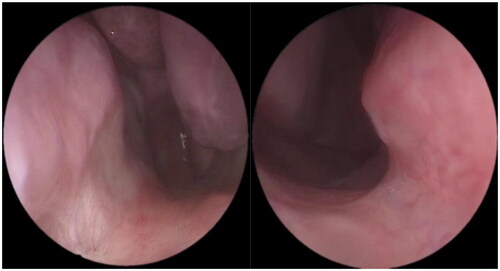
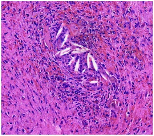
Figure 6. Pathological tissue shows a large number of inflammatory cell infiltrates and cholesterol crystals. Hematoxylin-eosin staining 100.