Figures & data
Figure 1. Preoperative findings. (A) Plain axial CT image shows calcified materials of various sizes (white arrow) in the left parotid gland. (B) Coronal T2-weighted MRI shows a hyperintense, lobulated lesion with multiple hypointense areas (white arrow) consistent with phleboliths in the left parotid gland.
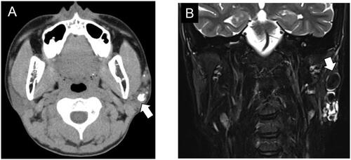
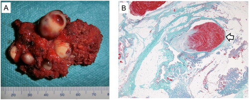
Figure 2. Excised specimen. (A) Macroscopic findings of the resected specimen are shown. Grossly, the parotid lesion is 54 mm in maximum diameter with multiple white stone-like objects. The largest object is 18 mm in size. (B) Elastic-Masson trichrome staining reveals nodular, irregular smooth muscle growth and disconnected elastic fibers in the abnormal vascular wall. Phleboliths (white arrow) are observed in the vascular lumen.