
Figures & data
Figure 1. Time course of computed tomography (CT). (a–c) Eight months prior to discovery of the mass, only coarse calcified lesions were detected in the right lobe of the thyroid (arrowheads), with no evidence of metastasis to the lungs or throughout the body. (d) and (e) Neck and chest CT before treatment indicating ∼4-cm hypoechoic mass in the right lobe of the thyroid gland, suggesting infiltration into the trachea and cervical esophagus and multiple nodules in lungs (arrowheads). (f) Positron emission tomography-CT before treatment demonstrating approximate accumulation in the right lobe of the thyroid and lung nodules, with no uptake detected in other organs. Although partial accumulation was noted in the small intestine, it was considered a false positive, as no lesions were identified on CT images. (g) and (h) Enlargement of tumor and infiltration into the trachea after treatment with doxorubicin. Slight shrinkage of pulmonary nodules (arrowheads) was observed. (i) and(j) Further progression of tumor and pulmonary nodules after treatment with pazopanib (arrowheads).
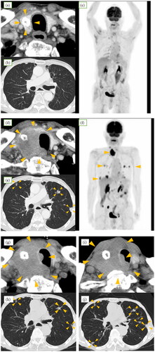
Figure 2. Pathology images. (a) Accumulation of atypical cells with enlarged nuclei, increased amounts of chromatin, and distinct nucleoli with poor binding on aspiration cytology (Papanicolaou staining). (b) Round to polygonal atypical cells with round nuclei and eosinophilic cytoplasm growing in sheet form around vessel (low magnification, hematoxylin & eosin staining). (c) The arrows in the image indicate increased mitotic activity (high magnification, hematoxylin & eosin staining). (d) Tumor cells immunohistochemically positive for smooth muscle actin. (e) Basement membranous material demonstrated by periodic acid-Schiff reaction. (f) Thyroid lesion (largest), forming 8.0 × 5.6 × 3.6-cm mass (yellow arrowheads) in contact with trachea (red arrowheads). (g) White mass which formed (yellow arrowheads) and infiltrated the trachea (red arrowheads).
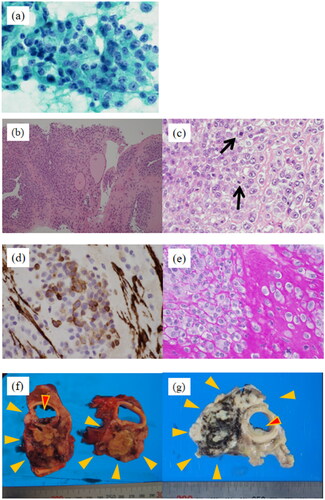
Table 1. Immunohistochemistry results.
Table 2. Case report of primary malignant glomus tumor of the thyroid gland.