Figures & data
Figure 1. Thalamita danae (Stimpson, 1858): (A) dorsal view, (B) ventral view, (C) frontal region of male, (D) basal antennal article, (E) outer surface of right cheliped of male, (F) carpus of chelipeds, (G) menus of chelipeds, (H) dactylus of natatory leg, (I) apical part of right G1 (dorsal view) and (J) apical part of right G1 (ventral view) and morphometric measurements.
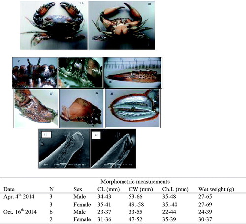
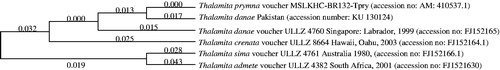
Figure 2. Molecular phylogenetic analysis of genus Thalamita by maximum-likelihood method Kimura 2-parameter model based on partial mitochondrial 16S rRNA gene (386 bp).