Figures & data
Figure 1. Ayun Musa thermal springs (A, B). Chara vulgaris population Linn. (C). C. vulgaris from the Springs of Moses cultivated in a glass vessel on a layer of fine sand, and showing nodes, internodes, and lateral branchlets (D).
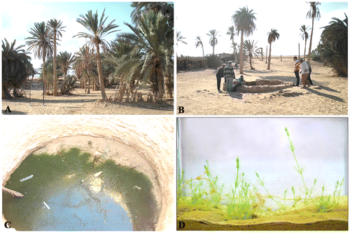
Table 1. Comparison of morphological and ecological characteristics of Chara vulgaris Linn. in this study and in reference literature.
Table 2. Key diagnostic features of Chara vulgaris from the Springs of Moses in freshly collected and cultured materials.
Figure (2–5). Some diagnostic characteristics of freshly sampled material of Chara vulgaris Linn. in this study. Scale bar = 1 cm (Fig. ); scale bar = 100 μm (Fig. ); scale bar = 1 mm (Fig. ); scale bar = 200 μm (Fig. ). (2) Macroscopic habitus of thallus showing long internodes. (3) Terminal branchlet cell. (4) Overview of branchlet node showing four well-developed bracteoles clearly much longer than oogonium. (5) Branchlet node with solitary dark-brown mature oogonium and absence of antheridium.
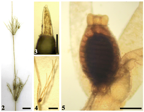
Figure (6–31). Diagnostic features of cultured material of Chara vulgaris Linn. in this study. Scale bar = 1 cm (Figs. ); scale bar = 100 μm (Figs. , , , ); scale bar = 200 μm (Figs. , ); Scale bar = 0.5 mm (Fig. ). (6, 7) Macroscopic habitus of Chara vulgaris thallus. (8) Part of thallus stem showing antheridia usually present at the newly formed branchlets at the stem apex (arrowhead), while oogonia are only still present on old branchlets (double arrowheads). (9–11) Whorls of branchlets usually curved inwards for the newly formed ones (arrowheads). (12–14) Nodes showing obtuse stipulodes in two closely placed rows (arrowheads). (15–18) Diplostichous, aulacanthous spine cells (arrowheads indicat spines present on the primary cortical rows, while double arrows depict larger secondary rows). (19) Elongated spine to the axis diameter in older stem. (20, 21) Terminal branchlet conical cells. (22) Fertile branchlet showing mature oogonia only and shedding antheridia. (23, 24) Fertile branchlet node with immature dark-green oogonium and solitary, spherical orange antheridium. (25, 26) Dark-black mature oogonia at branchlet node. (27, 28) Brown mature oospores with marked stripes. (29) Black mature oospore with pointed apex. (30–31) Internode cross-sections showing central siphon cells and cortical cells.
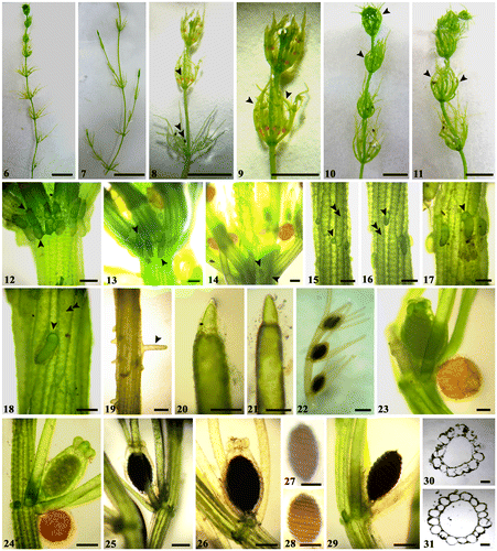
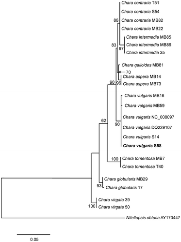
Figure 32. Maximum likelihood tree determined on the basis of partial matK gene sequences of 22 Chara samples. Outgroup = Nitellopsis obtusa (AY170447). The specimen from this study is marked in bold. Bootstrap values above 50 are included. The scale bar indicates 5% sequence divergence.
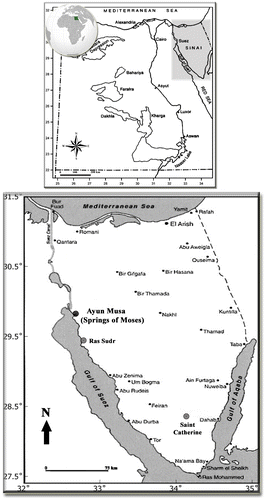
Map 1. Location of the sampling site in the Sinai Peninsula in Egypt.