Figures & data
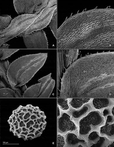
Figure 1. Selaginella germinans Valdespino & C. López. A. Section of upper surface of stem branch showing median and lateral leaves. B. Close-up of median leaf from main stem, upper surface; note, elongate and papillate idioblasts on both sides of the midrib (a), stomata along midrib (b) and on submarginal portion of lamina (c), and marginal, elongate and papillate idioblasts (d). C. Section of lower surface of main stem showing lateral leaves and portion of outer halves of median leaves. D. Close-up of proximal-acroscopic portion of lateral leaf (D1) and outer halve of median leaves (D2), lower surfaces; note, elongate and papillate idioblasts on both sides of the midrib (a), stomata along midrib (b) and on submarginal to submedial portions of lamina (c), and marginal, elongate and papillate idioblasts (d) on lateral leaf and submarginal stomata (e) and marginal, elongate and papillate idioblasts (f) on outer halve of median leaf. E. Megaspore, distal face; note close reticulate sculpturing pattern with high muri. F. Close-up of megaspore, distal face (same megaspore as in E); note high muri with perforate, sponge-like microstructure and rugulate and perforate microstructure on each reticulum lumen. A–F taken from holotype, Anderson et al. 7435 (holotype: NY).