
Figures & data
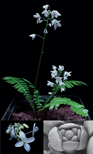
Figure 1. Floral morphologies in the Papaveraceae. (a) Eschscholzia californica (California poppy, Papaveroideae). (b) Pteridophyllum racemosum (pteridophylloideae). (c) Hypecoum imberbe (sicklefruit hypecoum, hypecoideae). (d) Lamprocapnos spectabilis (bleeding heart, Fumarioideae); floral diagrams are below the pictures with sepals colored in green, floral bract in dark blue, petals in grey, stamens in black, and the gynoecium as oval in the center. C, D referred to Sauquet et al. (Citation2015) and Hidalgo and Gleissberg (Citation2010); photos C, D, copyright free from Naturalist.org, C, by Quentin Groom, D, by Askalotl.
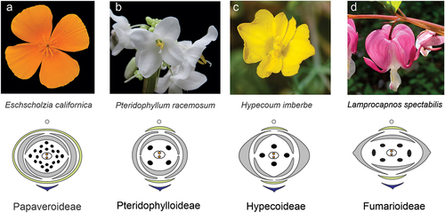
Figure 2. Pteridophyllum racemosum vegetative and floral morphology. (a) habitus of a flowering P. racemosum plant. (b) Pinnate leaf with leaflet numbers indicated. (c) scanning electron micrograph of the abaxial leaf epidermis including stomata. (d) SEM of multicellular adaxial trichomes, white lines indicated cell walls. (e) side view of the apical part of the inflorescence. (f) top view of a flower at anthesis. (g) view into the flower at anthesis with one petal removed. One sepal was falsely colored in yellow for clarity. Size bars correspond to (a, f) = 1 cm; (c) = 50 µm; (d) = 200 µm; (g) = 1 mm.
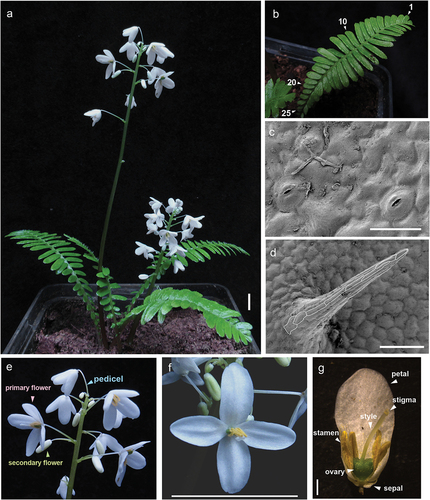
Figure 3. Histological sections showing different stages of P. racemosum floral development. (a) inflorescence meristems at the primary inflorescence apex. (b) longitudinal section through an inflorescence, subfigure assembled from multiple photos. (c) details of secondary inflorescence organization. (d) floral bud at stage 1 with sepal primordia initiated. (e) floral bud at stage 2, with petal primordia initiated. (f) floral bud at stage 2, when petals elongate. (g) floral bud at stage 3, when stamen primordia initiate. (h) floral bud at late stage 3, when stamen elongate and the gynoecium primordium becomes dome-shaped. (i) floral bud at stage 4, when the carpel walls initiates. (j) floral bud at stage 5, when carpel walls elongate and ovules initiate. (k) floral bud at stage 6, when carpels are fused at the apex. Longitudinal section subfigure assembled from two photos. (l) floral bud at stage 7, when the style elongates, integuments initiate, and male sporogenous tissue develops into pollen. (m) cross-section of floral bud in stage 7. Abbreviations: cw, carpel wall; fi, filament; fm, floral meristem; gp, gynoecium primordium; ii, inner integument; oi, outer integument; ov, ovule; p, petal; pim, primary inflorescence meristem; pp, petal primordium; s, sepal; sb, subtending bract; sim, secondary inflorescence meristem; sp, sepal primordia; stp, stamen primordium; st, stamen; sty, style; spt, sporogenous tissue; tt, tapetum tissue. Size bars correspond to (a, c, d, i-m) = 200 μm; (b) = 1 mm; (e-h) = 100 μm.
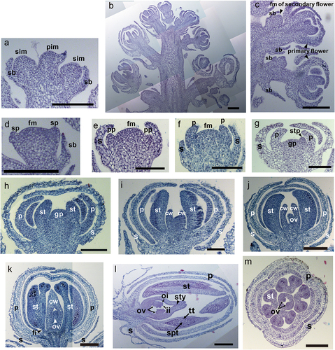
Table 1. Floral developmental stages of Pteridophyllum racemosum.
Figure 4. Scanning electron micrographs of P. racemosum floral development. (a) top view on an inflorescence with some buds removed. (b) floral bud at stage 1, encircled by a bract. (c) floral bud at stage 1, surrounded by a bract, with a subtending bract initiating. (d) floral bud at stage 3 with all floral organs initiated. (e) floral bud at late stage 4 with elongated outer petals. (f) floral bud at stage 6 with some petals and stamen removed to reveal the fused gynoecium. (g) Stigma and stamens grow up to the same height in late stage 6. (h) side view on the gynoecium with all other floral organs removed. The inset shows the stigmatic papillae at higher magnification. (i) side view on the ovary. (j) enlargement of the future dehiscence zone. (k) outer surface of the ovary wall. Abbreviations: b, bracts; dz, dehiscence zone; fm, floral meristem; gp, gynoecium primordia; ip, inner petal; o, ovary; op, outer petal; sb, subtending bract; sbm, subtending bract meristem; sti, stigma; sp, sepal primordia; s, sepal; st, stamen; sty, style. Scale bars (a) 1 mm; (b-d) 100 μm; (e,f,j) 200 μm; (g-i) 500 μm, enlarged fig in h: 50 μm; (k) 50 μm.

Supplemental Material
Download TIFF Image (29.2 MB)Data availability statement
Original data have been deposited here: http://dx.doi.org/10.22029/jlupub-18415.