Figures & data
Figure 1. Finite element model of the pelvis. (A) Whole pelvic model, (B) iliac bone.
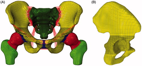
Table 1. The material properties of the pelvic tissues.
Figure 2. Fracture models. (A) Fracture component without any fixation systems, (B) the first fixation system: Linear two-lag screws configuration, (C) the second fixation system: single reconstruction plate with two-lag screws, (D) the third fixation system: single reconstruction plate with T-shaped mini-plates.
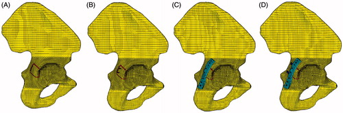
Figure 3. Pelvic model under four femur angle: (A) 0°, (B) 5°, (C) 10°, (D) 15°.
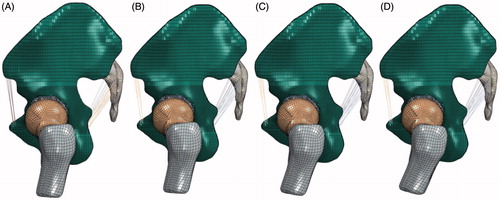
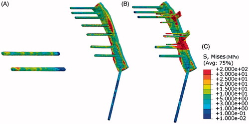
Figure 4. Displacement and stress distribution in two iliac bones. (A) Displacement distribution in iliac bone with large mesh size, (B) displacement distribution in iliac bone with small mesh size.
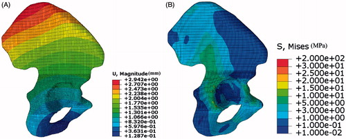
Table 2. The largest displacement in normal model, fracture model and three fixation systems under different load and boundary conditions.
Figure 5. The stress distribution in the iliac bone under different femur rotation angles with no fixation systems under 600N static vertical loads. (A) 0°, (B) 5°, (C) 10°, (D) 15°.
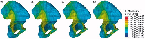
Figure 6. The stress distribution in the iliac bone under different femur rotation angles with no fixation systems under 1200N static vertical loads. (A) 0°, (B) 5°, (C) 10°, (D) 15°.
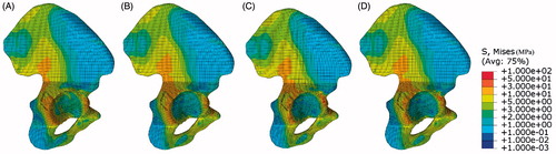
Figure 7. Stress distribution in iliac bones. (A) Stress distribution in normal model, (B) fracture component without any fixation systems, (C) the first fixation system, (D) the second fixation system, (E) the third fixation system.
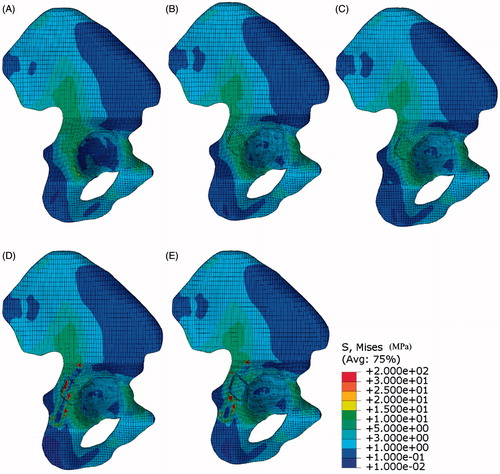
Figure 8. Von Mises stress along the limbus of acetabulum.
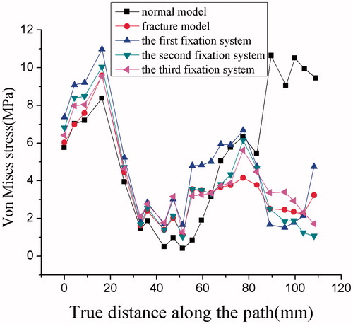
Figure 9. The relative displacement difference between different systems. (A) The outer line of the fracture component, (B) the inner line of the fracture component, (C) the relative displacement difference between different systems, (D) the relative displacement difference in the vertical direction component (i.e. the load direction) between different systems.
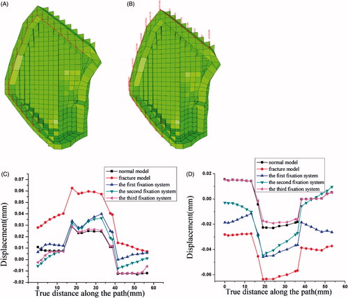
Figure 10. Stress distribution in fixation systems. (A) The first fixation system, (B) the second fixation system, (C) the third fixation system.