Figures & data
Figure 1. A 40-year-old female patient with the tumor of distal femur (#4). (a) Antero-posterior radiograph did not show the tumor clearly. (b, c) Axial and coronal views of computed tomography showed an obscure sclerotic lesion at the lateral epicondyle near the joint space. (d) Positron emission tomography showed positive FDG uptake in the tumor. (e, f) Axial and coronal views of the intraoperative 3D C-arm image. (g) Curettage was performed with the navigation system and reconstructed with artificial bone. (h) Postoperative computed tomography confirmed complete resection of the tumor.
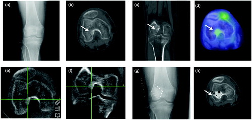
Table 1. Characteristics of the patients with tumor-induced osteomalacia.
Table 2. Preoperative and postoperative laboratory values.
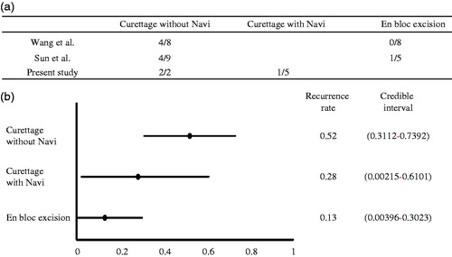
Figure 2. Comparison among three surgical methods. (a) Number of recurrence after using the three different methods for TIO in previous reports and in the present study. (b) Comparison of recurrence rate by Bayesian estimation. Navi: navigation system.