Figures & data
Figure 1. (a) CT of the Rando phantom in CT unit and details of TLD in (b) third section (c) seventh section and (d) the tenth section.
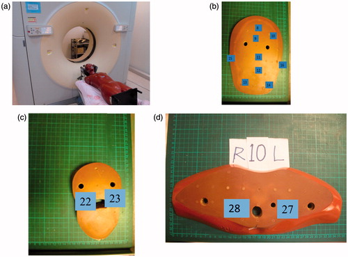
Figure 2. Scout view of two protocols obtained at Phillips Brillance 128-slice. (a) Axial scan: maxillae from external auditory meatus to parietal bone. (b) Helical scan: from mandible to parietal bone.
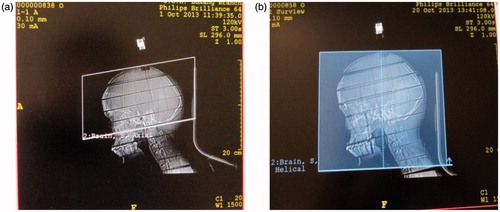
Table 1. Imaging parameters for two adopted protocols for brain CT routineexaminations.
Table 2. Weighting factor (WT) of organ or tissue recommended by ICRP 60 and 103 as well as number of TLD-100H imparted in Rando phantom.
Figure 3. Equivalent doses of organs or tissues (mSv) of the two protocols: The bar presents counting errors.
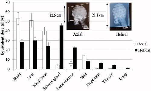
Table 3. listed organ or tissue doses (mSv) for 128-slice CT using axial and helical scan.
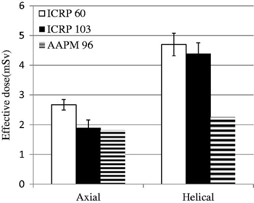
Figure 4. Calculation of E of the two protocols by different methods: TLD-100H approaches as recommend by ICRP 60 and 103;DLP method as recommend by AAPM 96. Error bar represented as counting errors.