Figures & data
Figure 1. 4-dimensional HyCoSy using SonoVue: (a) Both the fallopian tube and uterus were clearly visible. (b) contrast agent ringlike diffusion (arrow) around the ovary (asterisks). (c) the fallopian tubes (triangle) located on the side of ovary (asterisks) and the fluid collection (round) was an indirect sign of tubal patency.
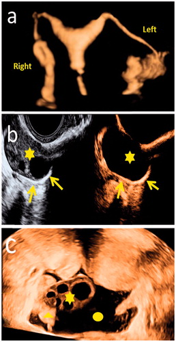
Table 1. Clinical qualities and the 4 D HyCoSy outcomes.
Table 2. Pain perception and side effects.
Table 3. The pain perception in patients with different degrees of patency.
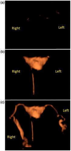
Figure 2. 4 D HyCoSy process: (a) contrast agent was not injected in the beginning. (b) contrast agent was injected into the catheter and next entered in the uterus. (c) Lastly, contrast agent developed in the bilateral fallopian tubes.