
Figures & data
Figure 1. Simplified results of femur model. (a) Original model, (b) 40% simplification, (c) 80% simplification, (d) 97.5% simplification and (e) Local amplification.
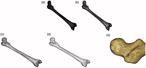
Figure 2. Mean and Hausdorff measurements of simplified model. (a) Mean distance of simplified model and (b) Hausdorffdistance of simplified model.
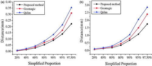
Figure 3. Mesh generation for femur. (a) Simplified model, (b) Partial model, (c) Surface mesh model, (d) Mesh refinement and (e) Body mesh model.
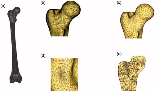
Figure 4. Mesh generation for vastus lateralis muscle. (a) Surface mesh model of vastus lateralis muscle and (b) Cutaway view of volume mesh of vastus lateralis.
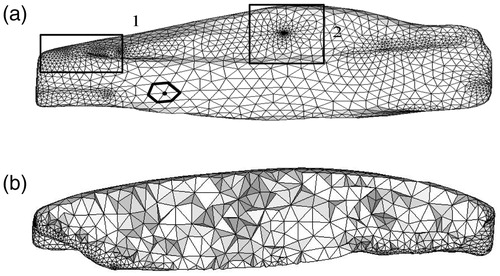
Figure 5. Assembled model of hip.
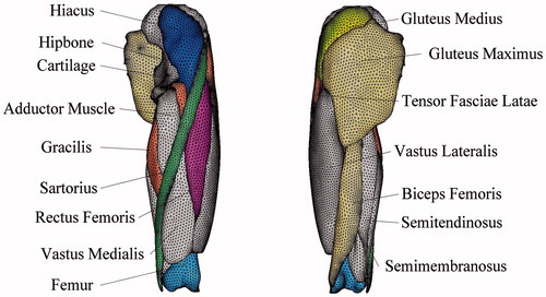
Figure 6. Partial graphs of femur mesh. (A1) Mesh generation result of proposed method, (A2) Mesh generation result of proposed method, (B1) Mesh generation result of hypermesh software and (B2) Mesh generation result of hypermesh software.
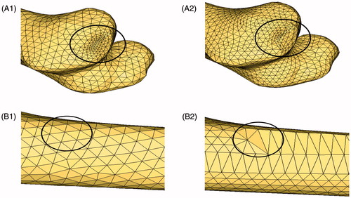
Table 1. Quality of surface triangular element.
Table 2. Mean and Haurdorff distances of surface mesh.
Figure 7. Quality analysis results for volume-mesh generation. (a) Quality analysis of volume-mesh generation for femur and (b) Quality analysis of volume-mesh generation for vastus lateralis muscle.
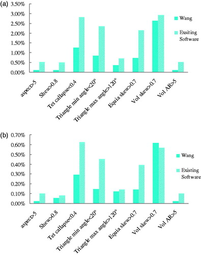
Figure 8. Tet collapse value distribution. (a) Tet collapse distribution of femur and (b) Tet collapse distribution of vastus lateralis muscle.
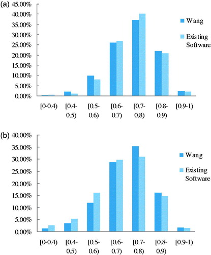
Table 3. Meshing efficiency analysis (unit: s).