
Figures & data
Figure 1. Pipeline for the proposed automatic surface-based registration method.
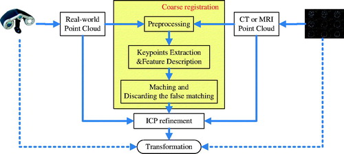
Figure 2. Illustration of the proposed registration process with a head phantom. Real-patient point cloud is green; CT point cloud is red. (a) Keypoint detection. The black dots indicate keypoints. (b) Keypoint correspondence. The blue lines indicate the correspondences. (c) Correspondences after discarding false matchings. (d) Coarse registration. (e) ICP refinement. (color online).
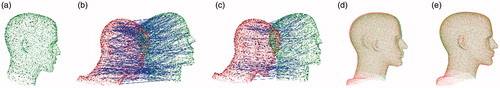
Figure 3. Facial region registration. The real-patient point cloud is green; the CT point cloud is red. (a) Only the facial region was scanned in the real-patient space. (b) Point cloud of the CT data. (c) Coarse registration. (d) ICP refinement. (e) An example of failed registration using a previously described method [Citation3]. The real-patient point cloud is golden; the CT point cloud is gray. (color online).
![Figure 3. Facial region registration. The real-patient point cloud is green; the CT point cloud is red. (a) Only the facial region was scanned in the real-patient space. (b) Point cloud of the CT data. (c) Coarse registration. (d) ICP refinement. (e) An example of failed registration using a previously described method [Citation3]. The real-patient point cloud is golden; the CT point cloud is gray. (color online).](/cms/asset/e83b12fb-0f84-4923-bf97-6ee8c96b19ef/icsu_a_1389411_f0003_c.jpg)
Figure 4. (a) TREs (mm) of each selected target point at different downsampling resolutions (the support radius was 18 mm). (b) TREs (mm) of each selected target point at different support radii for calculating feature descriptor (the edge length of the downsampling cube was 6 mm). (color online).
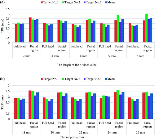
Figure 5. (a) Registration time against the length of divided cube for downsampling (the support radius was 18 mm). (b) Registration time against the support radius (the length of the downsampling divided cube was 6 mm). (color online).
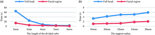
Figure 6. Clinical registration using the proposed method and a previously described 4PCS method [Citation3,Citation16]. I and II indicate patients one and two, respectively. (a) Point cloud from the real-patient surface. (b) Point cloud from MRI. (c) Full head registration. (d) Facial registration. (e) Failed facial registration using the 4PCS method. The real-patient point cloud is golden; MR image point cloud is gray. (color online).
![Figure 6. Clinical registration using the proposed method and a previously described 4PCS method [Citation3,Citation16]. I and II indicate patients one and two, respectively. (a) Point cloud from the real-patient surface. (b) Point cloud from MRI. (c) Full head registration. (d) Facial registration. (e) Failed facial registration using the 4PCS method. The real-patient point cloud is golden; MR image point cloud is gray. (color online).](/cms/asset/82e7d81a-560f-44cf-9b31-3748365ae34f/icsu_a_1389411_f0006_c.jpg)
Table 1. TREs (mm) of the clinical experiments. Numbers 1, 2, 3, and 4 are the numbers of four target points, and I and II indicate patients one and two, respectively.